


Key points from the article
- What mild proptosis may indicate: It can be benign or a sign of an underlying condition. If the underlying cause is not treated, more pronounced proptosis may lead to chronic dry eye, corneal ulceration, optic nerve compression and, in severe cases, vision loss.
- Five possible causes: High myopia associated with an elongated eyeball, congenital orbital anatomy, thyroid eye disease associated with Graves’ disease (Basedow’s disease), an orbital mass or inflammation, and lifestyle factors.
- Management: Depending on the cause and severity, management may involve regular monitoring, medical treatment or surgery. Urgent ophthalmic assessment is needed if warning signs develop.
- Outlook: Congenital or myopia-related proptosis does not resolve on its own, though surgery may improve it in selected cases. When proptosis is caused by an underlying condition, treating that condition, with input from an endocrinologist when appropriate, can help control symptoms and reduce the risk of complications.
1. What is mild proptosis? Is it dangerous?
What is mild proptosis?
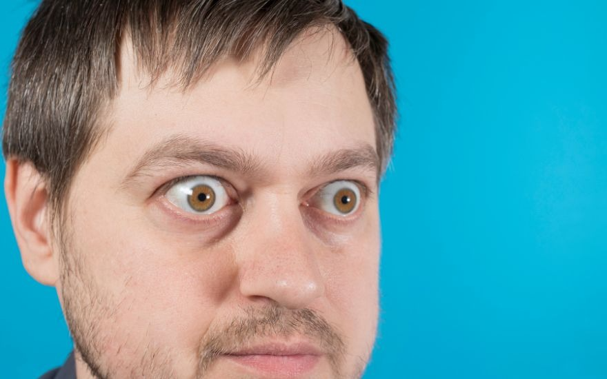
Proptosis is not a disease in itself. It is a sign of another medical condition or may result from congenital anatomy. It occurs when the tissues or muscles behind the eyeball become swollen or inflamed, pushing the eye forward. Mild proptosis is when the eyeball protrudes only slightly, making the white of the eye more visible, with mild retraction of the upper eyelid. At this stage, the person can still close their eyes fully, without the eyelids remaining open during sleep or experiencing dry, irritated corneas.[1]
The four grades of proptosis are as follows:
- Grade 1 (mild proptosis): 13–16 mm
- Grade 2: 17–20 mm
- Grade 3: 20–23 mm
- Grade 4: 24 mm
Is mild proptosis dangerous?
The level of risk depends on the underlying cause. Mild proptosis should not be ignored, as it may be an early sign of complications.[1]
- Benign cases (lower risk): If mild proptosis is due to congenital or hereditary orbital anatomy, or an elongated eyeball in people with high myopia, it is usually stable and does not progress over time. It mainly affects appearance and does not reduce vision.
- Cases caused by an underlying condition (requiring medical assessment and treatment): Mild proptosis may be an early sign of serious conditions such as hyperthyroidism (Basedow’s/Graves’ disease), an orbital mass or orbital cellulitis.
- Warning signs: Seek medical attention immediately if the proptosis progresses rapidly, affects only one eye or is accompanied by pain, redness, excessive tearing, double vision or blurred vision.
- Risks of delaying treatment: As proptosis worsens, the eyelids may no longer close fully, causing chronic dry eye, corneal inflammation and ulceration, optic nerve compression and even blindness. Anyone who notices an abnormal change should see an ophthalmologist and an endocrinologist promptly.

2. Symptoms of mild proptosis and early signs
Even when proptosis is mild, changes in the appearance of the eyeball may be noticeable when looking straight ahead, from an angle or from above. The most apparent sign is greater visibility of the white part of the eye (the sclera).
Because the eyeball protrudes, mild proptosis may make the eyes more sensitive to environmental factors. You may experience discomfort such as:
- Eye irritation and dryness: The eyes may feel sore or dry, or water continuously when exposed to bright light, wind or electronic screens.
- Restricted eye movement: You may feel discomfort when moving your eyes, find it difficult to blink freely or have trouble fully closing your eyelids during sleep.
- Reduced vision (if the condition progresses): As proptosis becomes more pronounced, vision may become blurred or double (diplopia), directly affecting everyday activities.
3. Five common causes of mild proptosis
Many people believe that wearing glasses is the only cause of proptosis. In fact, the condition may have five common causes:
3.1. High myopia (increased axial length)
In people with high myopia, the eyeball is often longer than normal. This elongation may make the eye appear more prominent, but this is not pathological proptosis; it is a change in the shape of the eyeball associated with myopia.2
In addition, strong prescription lenses can create an optical effect that makes the eyes look smaller. As a result, many people feel that their eyes look more prominent after removing their glasses, even though the actual position of the eyeballs has not changed.2

3.2. Congenital or hereditary anatomical features
Some people have prominent eyes because of the natural structure of their face and eye sockets. This normal anatomical feature may run in families and usually does not affect vision or eye health.1
If the eyes have appeared prominent since childhood, remained stable for many years and are not accompanied by symptoms such as eye pain, double vision, redness or reduced vision, the condition is generally not a cause for concern.1

3.3. An early sign of thyroid disease (Basedow’s/Graves’ disease)
Graves’ disease is one of the most common causes of proptosis in adults. It occurs when the immune system attacks the tissues and muscles around the eyeball, causing inflammation and swelling behind the eye that pushes the eyeball forward.3
In addition to proptosis, a person may experience a gritty sensation, dry or watery eyes, sensitivity to light, redness or pressure around the eyes. As the disease progresses, double vision or reduced vision may occur. If proptosis is accompanied by these symptoms, seek medical assessment promptly to have your thyroid function and eye health evaluated.3

3.4. Orbital infection or mass (a potentially serious cause)
In some cases, proptosis is caused by inflammation or an abnormal mass in the eye socket. When the tissues behind the eyeball swell or become compressed, the eyeball may be pushed forward.1
Typical signs include proptosis in one eye, asymmetry between the eyes and rapid progression. The person may also experience eye pain, eyelid swelling, double vision or reduced vision.1
If proptosis appears suddenly, especially in one eye only, seek an ophthalmic assessment promptly to identify the cause and receive timely treatment.1

3.5. Unhealthy lifestyle habits
Lack of sleep, prolonged stress and continuous use of electronic screens can overwork the eyes without giving them enough time to rest and recover. When looking at screens for long periods, people blink less often, which can destabilise the tear film and lead to dry, tired eyes. Sleep deprivation may also impair blood circulation and lymphatic drainage around the eyes, allowing fluid to build up and causing dark circles, under-eye bags and puffy eyelids.4
These changes may make the eyes appear larger or more prominent, even though the eyeballs are not actually protruding. In addition, a high-salt diet can increase fluid retention around the eye sockets and make the swelling more noticeable.5
This is usually temporary and may improve with adequate sleep, stress reduction, regular breaks from screen use and a balanced, lower-salt diet.

4. Severity assessment and management of mild proptosis
Management depends on the underlying cause and severity. Patients should monitor for accompanying symptoms to help determine the appropriate course of action.
4.1. Common presentation (home monitoring or cosmetic treatment)
Signs: The eyes have appeared prominent for many years, the appearance is stable and not progressing, and there is no pain, redness, swelling, double vision or reduced vision.1
Management:
- Seek an assessment for an accurate diagnosis: Even if the prominent eyes cause no pain or discomfort, patients should not self-diagnose or rely on home monitoring alone. An ophthalmologist should assess the eyes using a Hertel exophthalmometer, ocular ultrasound and an evaluation of the orbital structures. Periodic monitoring under a doctor’s guidance is appropriate only after confirming that the prominence is benign and related to congenital anatomy or high myopia.
- Consider cosmetic treatment if desired: Lifestyle changes or eye drops cannot alter the structure of the eyeball to reduce its prominence. Patients who wish to improve the appearance of prominent eyes or scleral show may consult an ophthalmic plastic surgeon about specialised procedures. Following an examination, the doctor may recommend options such as a non-incisional double-eyelid procedure, blepharoplasty, canthoplasty or other specialised procedures to improve facial balance while protecting visual function.

4.2. When to seek medical attention promptly
Signs: Proptosis accompanied by a gritty sensation, dry or irritated eyes, excessive tearing, redness or sensitivity to light. Some people may also feel pressure around the eyes or be unable to close their eyelids fully during sleep.3
Management: These symptoms may indicate damage to the ocular surface or proptosis associated with thyroid disease, such as Basedow’s/Graves’ disease. Patients should seek assessment at a specialist facility to determine the cause. Depending on the extent of the damage, the doctor may recommend:
- Corneal protection: Use artificial tears during the day and lubricating gel or eye ointment at night to keep the eyes moist and reduce the risk of corneal inflammation or ulceration due to incomplete eyelid closure.
- Inflammation control: Prescription eye drops or oral anti-inflammatory medicines, such as corticosteroids, to reduce inflammation and swelling in the tissues and muscles around the eye socket.
- Visual support: Wear sunglasses to reduce sensitivity to light, or use prism glasses if double vision (diplopia) has developed.
- Multidisciplinary consultation: If thyroid-related proptosis is suspected, an ophthalmologist may order blood tests (TSH, FT3 and FT4) and work with an endocrinologist to treat the underlying cause.

4.3. When to seek emergency care
Signs: Proptosis that appears suddenly, worsens rapidly over a short period or is asymmetric, affecting only one eye. Other symptoms may include pain around the eye socket, double vision, drooping eyelid, reduced vision, eye redness or swelling around the eye.1
Immediate action: This is an ophthalmic emergency and may be associated with serious conditions such as orbital cellulitis, orbital haemorrhage, a compressive orbital mass or optic nerve injury.
- What to do: Do not rub the eye, apply hot or cold compresses, or use eye drops without medical advice. Rest with the eye gently closed, avoid further irritation and ask someone to take you to a healthcare facility or the emergency department of a major hospital. Do not drive yourself.
- Medical assessment and treatment: At the hospital, doctors may order orbital imaging, such as a CT scan or MRI, to assess the location and extent of the damage and determine the appropriate treatment promptly, with the aim of protecting vision and preventing serious complications.
5. Can mild proptosis resolve on its own? Effective treatment options
Can mild proptosis resolve on its own?
The likelihood of improvement depends on the underlying cause. Proptosis due to congenital anatomy or high myopia does not resolve on its own. When proptosis is caused by thyroid eye disease, it may lessen once the underlying condition is controlled, but the eyeball rarely returns completely to its normal position.1
Treatment options:
- Proptosis related to myopia or congenital anatomy: There are currently no oral medicines, eye drops or exercises that can move the eyeball back into the eye socket. Contact lenses or refractive surgery may help patients avoid wearing glasses, but these options improve vision only and do not change the position of the eyeball. If proptosis affects appearance or daily activities, patients should consult an ophthalmologist about suitable options.
- Proptosis related to thyroid disease: Treatment focuses on controlling the thyroid condition and inflammation around the eye socket. Depending on the case, the doctor may prescribe medication and artificial tears to protect the ocular surface. If the underlying condition is stable but significant proptosis persists, surgery may be considered to restore eye function and improve appearance.

6. Home care and protection for mild proptosis
The following measures may help care for and protect the eyes at home:
- Manage dry eye: Proptosis exposes more of the ocular surface to the air, making it more prone to dryness. Use preservative-free artificial tears to keep the eyes moist and relieve a gritty or burning sensation.6
- Protect the eyes during sleep: Incomplete eyelid closure during sleep increases the risk of corneal dryness and ulceration.7 Use eye ointment at night or other protective measures as directed by your doctor.6
- Adjust daily habits: Limit prolonged, uninterrupted screen use to reduce eye strain and dryness. Wear UV-protective sunglasses outdoors to shield the eyes from dust and protect the ocular surface.6

7. Screening for causes of proptosis and personalised treatment at Hong Ngoc Eye Center
An incorrect diagnosis of the cause of proptosis, such as confusing myopia-related prominence with thyroid eye disease, may have serious consequences. Through its eye care services, Hong Ngoc Eye Center provides examination and diagnosis in accordance with international standards:
- One-to-one consultations and ongoing specialist support: Internationally trained ophthalmologists directly measure eye protrusion using a Hertel exophthalmometer and assess whether it is physiological or pathological. Patients are closely monitored and supported throughout treatment. This allows doctors to track the condition and adjust the treatment plan as needed, avoiding overlapping approaches and supporting optimal recovery.
- Advanced diagnostic testing: The Hospital uses modern equipment to identify the underlying cause of proptosis:
- IOLMaster: Doctors use the IOLMaster to measure axial length accurately and assess whether the prominence is associated with myopia.
- Ocular ultrasound and the NIDEK Mirante fundus imaging system: These tools help doctors detect orbital masses, enlargement of the extraocular muscles associated with Graves’ disease, or optic nerve damage.
- Multidisciplinary consultation and treatment of the underlying cause: Hong Ngoc Eye Center benefits from the Hospital’s multidisciplinary services. If thyroid-related proptosis is suspected, the ophthalmology team promptly consults with endocrinologists. This collaboration helps develop a treatment plan that addresses the underlying cause and prevents vision loss.
- Aesthetic treatment options: For patients with high myopia, Hong Ngoc Eye Center offers advanced refractive procedures, including SmartSight and phakic intraocular lens (ICL) implantation. These procedures can help patients avoid wearing glasses and may improve the cosmetic appearance of the eyes.

8. Frequently asked questions about mild proptosis
8.1. Is mild proptosis a cause for concern?
Mild proptosis is usually not dangerous if it has been present for a long time, remains stable and is not accompanied by pain or blurred vision. It is often related to congenital features or the structure of the eye socket. However, seek medical attention promptly if the proptosis is new, progresses rapidly or is accompanied by dry eyes, pain, double vision or reduced vision. These signs may indicate thyroid eye disease or an orbital condition.1
8.2. Can eye massage treat proptosis?
No. There is currently no medical evidence that massage can cure proptosis. Proptosis results from changes in the structure of the eyeball or eye socket, or from thyroid disease. Massage exercises cannot change the position of the eyeball. Massage may relieve eye strain, relax the muscles around the eyes and help with dry eye symptoms. If the eyes become increasingly prominent, consult an ophthalmologist to identify the cause.1
8.3. Does removing glasses make the eyes look less prominent in people with high myopia?
No. Removing glasses does not make the eyes less prominent or change the position of the eyeballs. Eye prominence depends on axial length, orbital anatomy or an underlying condition. Strong prescription lenses create an optical effect that makes the eyes look smaller. As a result, removing glasses may make the eyes seem more prominent, even though their actual position has not changed.8
8.4. Can mild proptosis be treated?
The outlook depends on the underlying cause. If proptosis is congenital or related to high myopia, its cause cannot be corrected with medication or exercises. If it is related to thyroid disease or orbital inflammation, appropriate treatment may reduce the degree of protrusion. However, the eyeball rarely returns completely to its normal position. If proptosis is new or progresses rapidly, see a doctor to identify the cause and determine the appropriate treatment.1
Mild proptosis is a clinical sign that should be monitored and accurately assessed by a specialist. Whether it is related to a benign anatomical feature or a more complex medical condition, identifying the cause and intervening promptly are essential to protecting vision and improving appearance. Do not ignore abnormal forward protrusion of the eyeball or attempt to treat it without medical advice.
In-depth ophthalmic examination and diagnosis at Hong Ngoc Hospital
With an experienced ophthalmology team and modern diagnostic equipment, including the IOLMaster and NIDEK Mirante fundus imaging system, Hong Ngoc Eye Center provides in-depth screening and personalised treatment plans for proptosis in accordance with medical standards
If you have any eye concerns, contact Hong Ngoc General Hospital on 0916 987 366 for timely assistance.
Note: The information in this article is for reference only and is not a substitute for professional medical advice or diagnosis. Each person’s eye condition and anatomy are different. Patients should undergo a thorough examination and individual assessment by an ophthalmologist before deciding on a treatment plan.
References:
- Proptosis (bulging eyes). (2021, May 15). Cleveland Clinic. https://my.clevelandclinic.org/health/diseases/proptosis-bulging-eyes
- Kelley, S., O.D, & M.S. (n.d.). Is nearsightedness due to the eyeball being too long? All About Vision. Retrieved July 30, 2026, from https://www.allaboutvision.com/conditions/myopia/length-of-eye/
- Graves’ disease. (n.d.). Mayo Clinic. Retrieved July 30, 2026, from https://www.mayoclinic.org/diseases-conditions/graves-disease/symptoms-causes/syc-20356240
- Sundelin, T., Lekander, M., Kecklund, G., Van Someren, E. J. W., Olsson, A., & Axelsson, J. (2013). Cues of fatigue: effects of sleep deprivation on facial appearance. Sleep, 36(9), 1355–1360. https://doi.org/10.5665/sleep.2964
- Bags under eyes. (n.d.). Mayo Clinic. Retrieved July 30, 2026, from https://www.mayoclinic.org/diseases-conditions/bags-under-eyes/symptoms-causes/syc-20369927
- Dry eyes. (n.d.). Mayo Clinic. Retrieved July 30, 2026, from https://www.mayoclinic.org/diseases-conditions/dry-eyes/symptoms-causes/syc-20371863
- Lagophthalmos. (2022, November 7). Cleveland Clinic. https://my.clevelandclinic.org/health/diseases/24413-lagophthalmos
- Briceno, C. A. (2024, November 7). Nearsightedness: What is myopia? American Academy of Ophthalmology. https://www.aao.org/eye-health/diseases/myopia-nearsightedness