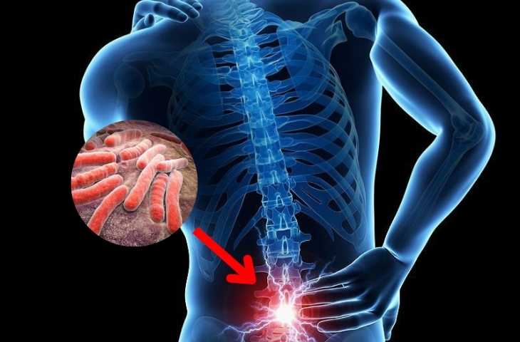
Spinal tuberculosis is an infectious inflammatory condition of the vertebrae and intervertebral discs caused by Mycobacterium tuberculosis, which may lead to spinal cord compression and subsequent motor impairment.
How can spinal tuberculosis be detected, and what treatment options are available? Let us explore.
What is spinal tuberculosis?
Spinal tuberculosis, also known as Pott’s disease, is a form of extrapulmonary tuberculosis in which Mycobacterium tuberculosis infects the vertebrae and intervertebral discs. This acid fast bacillus, measuring approximately 3–5 μm in length, can persist in the human body for several months.
Once it invades the spine, the bacteria proliferate and progressively destroy the vertebral bodies, leading to vertebral collapse and potential spinal instability. Spinal tuberculosis is a serious condition that requires early diagnosis and prompt treatment.
Risk factors for spinal tuberculosis
Spinal tuberculosis can affect individuals of all ages; however, certain groups are at higher risk, including:
- Individuals with a history of pulmonary or skeletal tuberculosis
- Those with close contact with tuberculosis patients, particularly within the household
- Immunocompromised individuals, including those with chronic diseases, malignancies, or HIV/AIDS
- Males, who have a higher incidence compared to females
Etiology of spinal tuberculosis
Mycobacterium tuberculosis typically enters the body via the respiratory tract, initially affecting the lungs. Through hematogenous or lymphatic dissemination, the bacteria can spread to other organs, including the spine within the musculoskeletal system.
The main routes of transmission include:
- Direct contact with individuals infected with tuberculosis
- Entry of the bacteria through open wounds or skin abrasions
- Vertical transmission from mother to child

Common clinical manifestations of spinal tuberculosis
Spinal tuberculosis often progresses insidiously, with symptoms typically appearing at advanced stages. Common manifestations include:
- Localized spinal pain: Initially presenting as dull, persistent pain at the affected vertebral level (cervical, thoracic, or lumbar), often worsening at night or in the early morning. As the disease progresses, pain becomes more severe, limits mobility, and may radiate along nerve roots, sometimes causing lower limb spasms.
- Progressive muscle wasting of the lower limbs: Atrophy of the legs, particularly in the calves or anterolateral lower legs, due to spinal cord compression, leading to impaired mobility.
- Paravertebral or psoas abscess formation: A soft mass (cold abscess) may develop and track along anatomical pathways to the groin, buttocks, thighs, or gluteal region. Large abscesses may rupture, causing secondary infection, inflammation, and pain.
- Paraplegia: Loss of motor function in both lower limbs, commonly resulting from spinal cord compression and impaired blood supply, particularly in lower thoracic spinal involvement.
Diagnostic approaches for spinal tuberculosis
Based on clinical presentation, imaging findings, and advanced laboratory investigations, spinal tuberculosis can be diagnosed from early stages. Key diagnostic methods include:
- Spine X ray: May reveal vertebral body destruction, narrowing of intervertebral disc spaces, and cavitary lesions caused by Mycobacterium tuberculosis. However, X ray often detects the disease only at advanced stages when structural damage is significant.
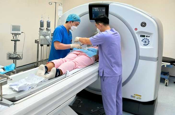
- Magnetic resonance imaging (MRI) or computed tomography (CT): These advanced imaging modalities allow earlier and more accurate detection, including disc space narrowing, bilateral vertebral body destruction, and the presence of paravertebral or psoas abscesses. MRI is particularly useful for assessing soft tissue involvement and spinal cord compression.
- Differential diagnosis: Spinal tuberculosis is characterized by bone destruction without significant reactive sclerosis or periosteal thickening, and often involves both vertebral bodies and intervertebral discs. In contrast, bone malignancies typically affect the vertebral bodies while sparing the intervertebral discs.
- Bone biopsy: Involves obtaining tissue samples from the affected vertebrae for microbiological and histopathological analysis, enabling definitive identification of Mycobacterium tuberculosis. This is especially indicated when imaging findings are inconclusive.
- Additional laboratory tests: Supporting investigations include a positive Mantoux test, elevated erythrocyte sedimentation rate (ESR), GeneXpert MTB/RIF assay, and Quantiferon TB Gold Plus test.
At the Department of Musculoskeletal Medicine, Hong Ngoc General Hospital, spinal tuberculosis is diagnosed using a range of advanced technologies, supported by modern equipment systems:
SIGNA Prime MRI system with AIR reconstruction technology enables early detection of abscesses and vertebral or intervertebral disc lesions
Reader may also be interested in:
- Is cervical spondylosis dangerous? What are the most effective treatment options?
- Symptoms of cervical spondylosis and effective prevention strategies
128 slice CT scanner (USA) reduces radiation exposure by up to 82% while providing high resolution imaging of the cervical, thoracic, and lumbar spine, improving efficiency and patient safety
CT guided biopsy with image intensifier ensures precise localization, trajectory, and depth of tissue sampling, minimizing invasiveness and enhancing diagnostic accuracy
Notably, the medical team includes physicians with experience at leading domestic and international institutions such as Bach Mai Hospital, Friendship Hospital, and Institut Pasteur (France), ensuring appropriate diagnostic approaches and timely treatment for patients.
Management of spinal tuberculosis
The treatment of spinal tuberculosis requires strict adherence to medical guidance and long term compliance:
- Early stage: When the infection is localized, conservative management is indicated, including anti tuberculosis pharmacotherapy combined with physiotherapy and spinal immobilization. Anti tuberculosis drugs eradicate and inhibit bacterial growth, while physiotherapy helps relieve pain, reduce inflammation and edema at affected vertebral levels, and prevent muscle atrophy and joint stiffness.
- Advanced stage: In cases where the infection progresses with abscess formation and risk of spinal cord or visceral compression, surgical intervention may be required, along with strict adherence to a comprehensive treatment regimen prescribed by specialists.
In addition, to enhance immune function, patients may consider multivitamin supplementation and maintain adequate nutrition through a balanced daily diet.
Note: The information provided in this article by Hong Ngoc General Hospital is for reference purposes only and does not replace professional medical diagnosis or treatment. Patients should not self medicate. For accurate diagnosis and appropriate management, individuals are advised to visit a healthcare facility for direct examination, diagnosis, and consultation with qualified physicians.
Follow the official fanpage of Hong Ngoc General Hospital for more health updates and useful information:https://www.facebook.com/BenhvienHongNgoc