


The cornea is the transparent outermost layer of the eye. It acts like a lens, focusing light so that we can see clearly. When it is severely damaged and permanent scarring or opacity develops, light can no longer pass through properly, resulting in vision loss.
Early and accurate identification of the causes of corneal blindness is an important first step in preventing risk. It also helps patients access timely medical intervention to preserve and restore vision. This article examines the causes in detail and explores specialised treatment options.
Key points in this article
- Why corneal damage is serious: Any corneal injury can progress rapidly, may leave permanent scarring or opacity, and can lead to reduced vision or blindness if not treated appropriately.
- Four main groups of causes: Corneal blindness is most commonly associated with infections (bacterial, viral or fungal); injuries and burns (mechanical, chemical or thermal); hereditary or degenerative conditions, such as keratoconus; and vitamin A deficiency or systemic disease.
- Red flag symptoms: Seek emergency ophthalmic care for severe eye pain, a gritty sensation, redness, light sensitivity, persistent tearing or sudden vision loss.
- Treatment and vision restoration: The key principle is that time is of the essence. Early injuries are generally treated with medication. When the cornea has developed scarring or severe distortion, corneal transplantation—penetrating, lamellar or endothelial keratoplasty—may be the most effective medical option for restoring vision.
- Risk prevention: Do not use eye drops without medical advice, especially those containing corticosteroids, if your eye is red or scratched. Wear protective eyewear when working and follow medical guidance on contact lens hygiene to protect the ocular surface.
1. Causes of corneal damage that can lead to blindness
The invasion of corneal tissue by harmful microorganisms is a leading cause of acute keratitis and corneal ulcers. The main types include:
1.1. Infectious keratitis and corneal ulcers
Infection is a common cause of corneal scarring and blindness, but it can be prevented. When the protective corneal epithelium is damaged by an injury or foreign body, or through improper contact lens use, bacteria, viruses, fungi or parasites can enter the corneal stroma and cause ulceration. Without prompt diagnosis and treatment, the affected area may develop permanent opacity and scarring, leading to reduced vision or blindness.1
- Bacterial infection: Bacterial keratitis often develops after a corneal injury, particularly among agricultural and mechanical workers, or as a result of improper contact lens use, such as wearing lenses overnight or failing to clean lenses and their cases as directed. Some pathogens, including Pseudomonas aeruginosa, can progress rapidly and cause severe corneal damage if not treated promptly.2
- Viral infection: Herpes simplex virus (HSV) is a common cause of viral keratitis. Typical symptoms include eye redness, pain, tearing, light sensitivity and blurred vision. After the initial infection, the virus can remain dormant in a sensory nerve ganglion and reactivate when the immune system is weakened, during periods of stress or in the presence of other illnesses. Recurrent episodes can cause corneal scarring and vision loss and, in severe cases, blindness.3
- Fungal and parasitic infections: Fungal keratitis often follows an eye injury involving plant material, such as a branch, rice leaf or thorn striking the eye. It is particularly common in tropical countries. Acanthamoeba keratitis, meanwhile, is often associated with improper contact lens use, including exposure to tap water, swimming pools or shower water while wearing lenses. Both conditions are difficult to treat, often require prolonged treatment and can cause severe corneal scarring if diagnosis is delayed.4

1.2. Eye injuries and corneal burns
Blunt eye trauma, chemical burns and thermal burns can cause severe corneal damage. The extent of the damage depends on the type of agent, duration of exposure and initial management. Without prompt emergency care and treatment, these injuries may cause corneal scarring, vision loss or blindness.5
- Blunt and penetrating injuries: Workplace, road traffic or household accidents can cause corneal abrasions, lacerations or perforations. Open wounds can allow bacteria to enter the eye, leading to keratitis, corneal ulcers or endophthalmitis. Severe injuries may leave permanent scarring, cause significant vision loss or even require surgical removal of the eye if it cannot be salvaged.5
- Chemical burns: If the eye is exposed to a chemical, it should be irrigated immediately before the person seeks medical care. Acid burns usually cause proteins on the surface to coagulate, forming a barrier that limits deeper penetration of the chemical into the eye. In contrast, alkalis such as ammonia, lime, cement and alkaline cleaning products can destroy cell membranes and penetrate rapidly and deeply into the eye. Alkali burns can cause necrosis of the cornea and conjunctiva, increasing the risk of vision loss or blindness if treatment is delayed.6
- Thermal and radiation burns: High temperatures, welding sparks or intense ultraviolet (UV) radiation can damage the corneal epithelium. Symptoms often include severe pain, a gritty sensation, excessive tearing, light sensitivity and difficulty opening the eyes. Most mild epithelial injuries can heal with appropriate medical treatment. However, severe burns or prolonged exposure may still result in corneal scarring and vision loss.7
1.3. Inherited and degenerative conditions (non-inflammatory)
Not all corneal damage is caused by inflammation or injury. Some inherited or progressive degenerative corneal conditions can distort the cornea or gradually reduce its transparency. Without early diagnosis and treatment, they may cause severe vision loss and, in some cases, require corneal transplantation to preserve vision.
- Keratoconus: The cornea gradually thins and bulges forward into a cone shape. This change in corneal shape can cause unusual progression of myopia and astigmatism, with vision becoming increasingly blurred and difficult to correct with spectacles. In advanced cases, corneal scarring or acute corneal hydrops may develop, potentially leading to permanent blindness.8
- Corneal dystrophies: This group of inherited conditions gradually affects the structure of the cornea. Fluid may build up in the cornea, causing swelling and loss of its natural transparency, which reduces vision. As the condition progresses, tiny blisters may form on the ocular surface, causing a persistent gritty sensation and severe pain.9

1.4. Nutritional deficiencies and systemic conditions
Nutritional deficiencies and certain systemic conditions can weaken the cornea, delay healing and increase the risk of corneal ulcers and scarring.10
- Vitamin A deficiency: Vitamin A is essential for maintaining a healthy cornea. Prolonged deficiency can cause severe dryness and weaken the surface cells, making the cornea more prone to ulceration. In advanced cases, the cornea may soften (keratomalacia), become necrotic or even perforate. Without timely medical intervention, this can lead to permanent blindness.
- Systemic and autoimmune conditions: Some autoimmune diseases, such as Sjögren’s syndrome, rheumatoid arthritis and lupus, can cause chronic dry eye and severe damage to the ocular surface. Diabetes, a common systemic condition, can also impair immune function and slow wound healing in the eye. If these conditions are not well controlled, they can increase the risk of inflammation, corneal ulceration and erosion, and permanent scarring.

2. Warning signs of serious corneal damage requiring emergency care
The cornea has a very dense network of nerves, so any injury to it can cause severe pain and directly threaten vision in a very short time. If you experience any of the following red flag symptoms, do not ignore them. Seek emergency ophthalmic care immediately. Delayed treatment can lead to permanent blindness, particularly if you have a history of eye trauma or wear contact lenses.11
- Sudden decline in vision: Vision may become blurred rapidly, as though a fog or film has come over the eyes. The degree of vision loss depends on the location and severity of the corneal damage.11
- Severe eye pain and a gritty sensation: The eye may hurt intensely and feel as though there is a foreign body in it, with pain worsening when you blink or move your eye. These are typical symptoms of a corneal abrasion or ulcer.12
- Redness, bleeding and light sensitivity: The white of the eye may become intensely red, with prominent blood vessels or even patches of bleeding around the edge of the iris. The eye may also become extremely sensitive to light, causing sharp pain and making it difficult to open normally.13
- Persistent tearing: Excessive tearing is a natural reflex that helps protect the corneal surface when it is irritated or injured. It often occurs alongside pain and a gritty sensation.12
- White spots or an ulcer on the cornea: White spots, cloudy patches or an ulcer on the black part of the eye (the cornea) may indicate bacterial, fungal or other infectious keratitis. This is an ophthalmic emergency requiring urgent medical treatment to prevent corneal scarring or permanent vision loss.11
3. Treatment options and the potential to restore vision
Treatment and the potential for vision recovery depend on the location, cause and actual severity of the corneal damage. Based on diagnostic imaging results, the doctor will recommend an appropriate intervention.
3.1. Conservative medical treatment: when is medication sufficient?
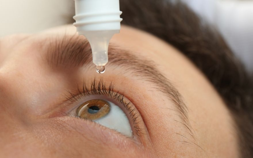
Medication is the preferred treatment for early-stage damage, mild corneal ulcers or superficial abrasions, when the corneal structure has not been severely damaged. The main goals are to eliminate the causative pathogen and prevent corneal scarring and opacity.14
Depending on the cause, the doctor may prescribe specific medicines, applied topically or taken orally, such as antibiotics for bacterial infections, antifungal medicines or antiviral medicines for herpes simplex. For damage caused by autoimmune disease, anti-inflammatory or immunosuppressive medicines may be used to control inflammation and support corneal healing.14
3.2. Corneal transplantation: when is surgery necessary?
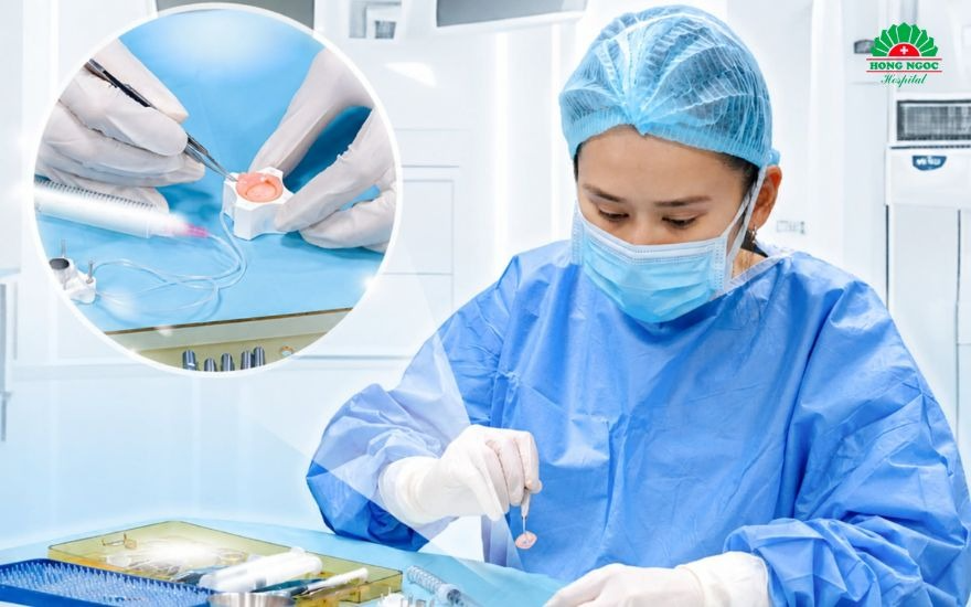
When damage is severe, the cornea has permanent scarring or opacity, perforation, severe distortion (such as keratoconus) or complete loss of transparency, and vision is significantly impaired despite medical treatment, corneal transplantation may be the best option for restoring vision.15
Modern medicine offers more than one conventional approach, allowing surgeons to target the affected layers of the cornea. The technique is selected according to which layer is damaged.15
- Penetrating keratoplasty (PK): Replaces the full thickness of the cornea, from the outer epithelium to the innermost endothelium, with healthy donor tissue. It is used for very deep scars or damage involving the full thickness of the cornea.
- Anterior lamellar keratoplasty (ALK/DALK): Removes and replaces only the diseased anterior layers while preserving the healthy underlying endothelium. This technique can significantly reduce the risk of graft rejection.
- Endothelial keratoplasty (DSAEK/DMEK): Replaces only the thin endothelial layer on the inner surface of the cornea. It is commonly used to treat Fuchs endothelial corneal dystrophy. The procedure is less invasive and uses a very small incision, which can allow vision to recover more quickly.
- Femtosecond laser-assisted corneal transplantation: Instead of using a conventional mechanical surgical blade, the surgeon uses a highly precise femtosecond laser to create the corneal incisions. This technology enables a precise fit between the donor graft and the recipient’s eye, promotes faster healing and may reduce the risk of postoperative astigmatism.
3.3. The potential for vision recovery after corneal damage
Advances in modern ophthalmology, particularly microsurgical techniques and specialised corneal transplantation, have given many patients who once faced the risk of permanent blindness a chance to regain their sight.
However, the degree of vision recovery varies from person to person and depends on several key factors: the severity of the initial injury, how soon medical treatment begins, the function of other structures within the eye, such as the retina and optic nerve, and the person’s overall health.
In ophthalmology, the guiding principle is that time is of the essence: the sooner an injury is detected and treated appropriately, the greater the chance of preserving vision.
4. Effective measures to prevent corneal damage
Taking proactive steps to protect the eyes at work and in daily life is essential to reduce the risk of ocular surface damage and prevent vision loss. Follow these key preventive measures:
- Wear protective eyewear when doing work that carries a risk of eye injury: Always wear safety glasses that meet relevant standards when working around dust, metal fragments, chemicals, cutting or grinding equipment, or when taking part in activities that could injure the eyes. Protective eyewear can significantly reduce the risk of foreign bodies entering the eye and damaging the cornea.16
- Use contact lenses correctly: Do not sleep in contact lenses unless your doctor has prescribed lenses for overnight wear. Do not wear them while swimming or showering, rinse them with neither tap water nor other non-sterile water, wash your hands before inserting or removing them, and replace them as directed. Improper contact lens use increases the risk of severe corneal ulcers.17
- Protect your eyes from ultraviolet (UV) rays: Wear sunglasses that block 99–100% of UVA and UVB rays outdoors, especially in places with high UV exposure, such as beaches or mountainous areas. This helps reduce UV damage to the cornea and other eye structures.16
- Never rub your eyes if there is a foreign body: If dust or another object gets into your eye, do not rub it, as this may scratch the corneal epithelium. A safe first step is to blink repeatedly or rinse the eye with sterile saline to flush the object out. If it remains lodged in the eye, or if you have persistent pain, redness or blurred vision, seek medical care promptly so a doctor can remove it safely.18
- Maintain a balanced diet: Vitamin A plays an important role in maintaining a healthy corneal epithelium and preventing damage caused by vitamin A deficiency and dry eye. Increasing omega-3 fatty acids in your diet may also help protect the ocular surface and significantly improve clinical symptoms of dry eye.19

5. Specialist screening and diagnosis of corneal damage at Hong Ngoc Eye Center
Some patients with a corneal abrasion or mild eye redness buy corticosteroid-containing eye drops from a pharmacy without consulting a doctor. This can worsen an infection and lead to serious complications, including corneal ulcers, perforation and permanent vision loss. Any unusual eye symptoms should therefore be assessed by a reputable ophthalmology service.
5.1. One-to-one care from leading ophthalmology specialists
At Hong Ngoc Eye Center, patients are examined and diagnosed, and their treatment plans are developed by experienced ophthalmologists who have worked at leading healthcare facilities and received international training:
- Dr Vu Thi Tue Khanh: An ophthalmology specialist with 30 years of experience, with expertise in corneal disease, ocular surface disorders and corneal transplantation. She has worked at the Vietnam National Eye Hospital and LV Prasad Eye Institute in India, and has performed thousands of corneal transplants and corneal epithelial stem cell transplants.
- Dr Dinh Thi Hoang Anh: Head of the Eye Department at the Phuc Truong Minh campus. She holds 10 patents for microsurgical techniques recognised by Vietnam’s Ministry of Health and the Russian government, including PDEK endothelial keratoplasty. She has also undertaken extensive study and research at the A. I. Yevdokimov Moscow State University of Medicine and Dentistry in Russia.
- International case conferences: The hospital regularly holds in-person consultations with Prof. Dr Kalinnikov Yuri, a professor at a Russian medical university, to determine the optimal approach for complex cases of corneal damage.

5.2. Personalised diagnostic and medical treatment pathway
At Hong Ngoc Eye Center, diagnosis and treatment follow a medically appropriate process designed to ensure accuracy and effective care for each patient:
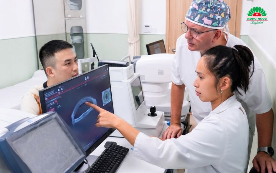
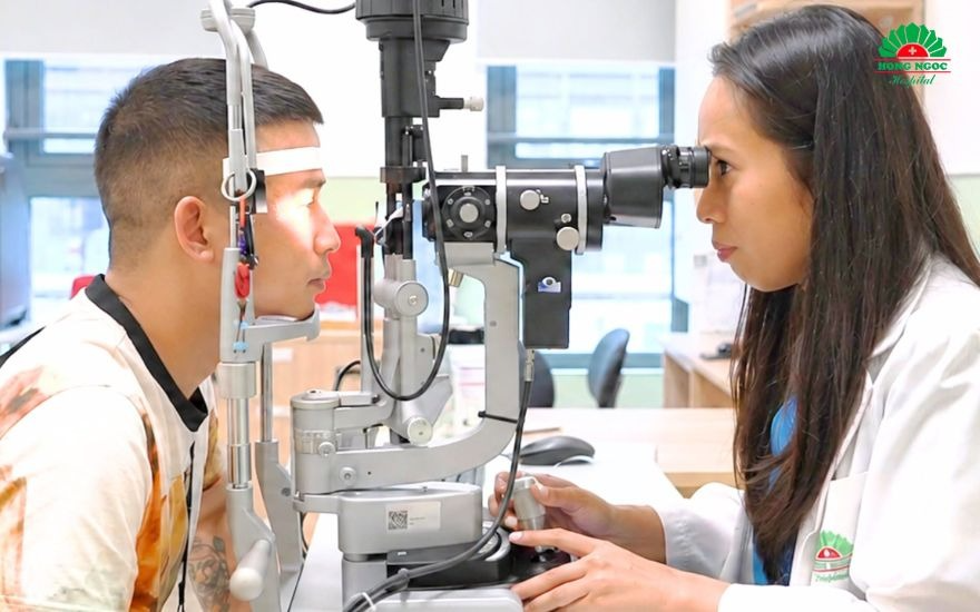
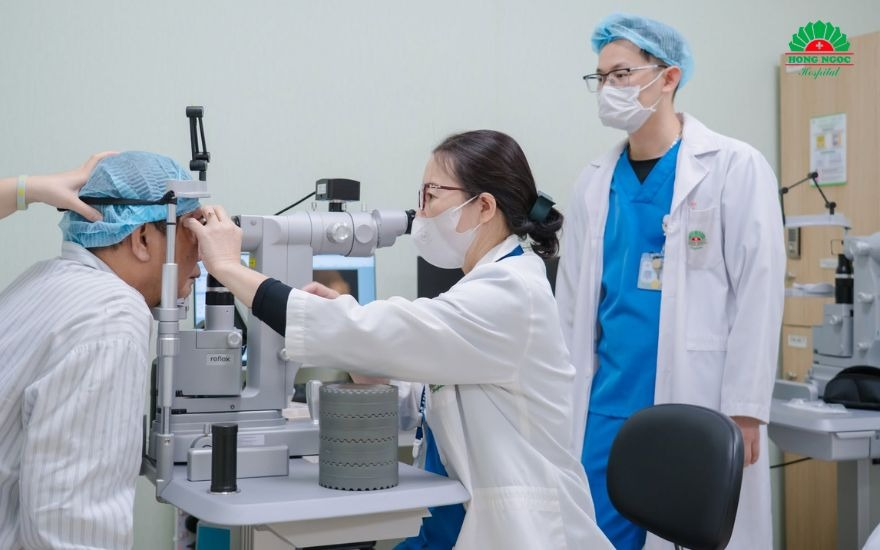
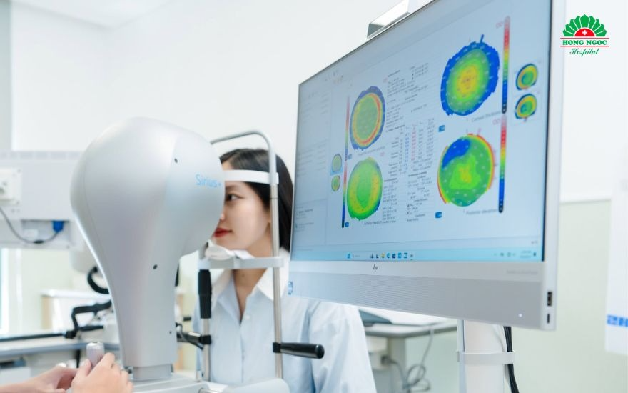
- Advanced diagnostic testing: The doctor uses fluorescein dye and slit-lamp biomicroscopy, as well as the Schwind Sirius+ corneal topography system, to assess corneal thickness and structure in detail and detect underlying conditions such as keratoconus or corneal degeneration at an early stage.
- A clear treatment plan: Depending on the cause, whether bacterial, viral or fungal infection, or injury, the doctor will prescribe specific medical treatment. For severe cases resulting in permanent corneal scarring or opacity, patients receive specialist advice on corneal transplantation options.
5.3. Corneal transplantation to international standards
Hong Ngoc Eye Center is one of the few private healthcare providers in Vietnam with the expertise to perform complex corneal transplantation procedures:
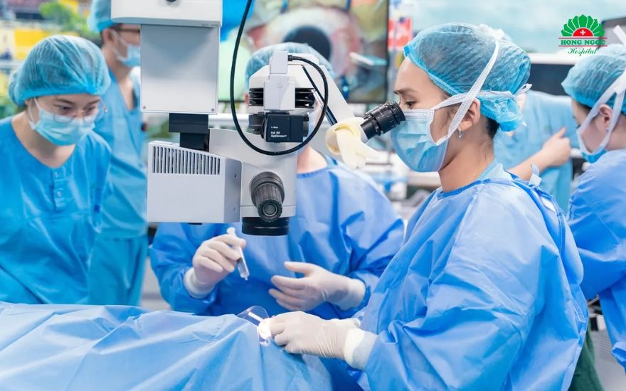
- Use of modern transplant technology: The center performs advanced microsurgical techniques, including selective lamellar keratoplasty using laser technology, and incorporates proprietary innovations from Russia.
- High-level collaboration: Hong Ngoc is a coordinating member of the Vietnam Cornea Bank, which is part of the Vietnam National Eye Hospital, supporting access to legally sourced, safe donor tissue and comprehensive treatment. The hospital is also a strategic partner of the MNTK Fyodorov Eye Microsurgery Complex in Russia for technical transfer and remote case consultations.
- Surgical safety assurance: The hospital’s healthcare quality is recognised through international ACHSI accreditation, and it is a Global Surgical Training Centre of the Royal College of Surgeons of England (RCS). Corneal transplants are performed in operating theatres designed to meet UK HBN standards, with stringent infection prevention and control procedures.
- Multidisciplinary consultations: As a multispecialty hospital, Hong Ngoc can screen, treat and closely manage patients’ systemic conditions before recommending corneal transplantation. This supports higher success rates and helps reduce the risk of graft rejection.
6. Frequently asked questions (FAQs) about corneal damage
6.1. Can a mild corneal abrasion heal on its own?
Yes. Most mild corneal abrasions heal on their own within 24–72 hours, as the corneal epithelium regenerates rapidly. However, you should still see a doctor to rule out a retained foreign body, infection or deeper injury. During this time, avoid rubbing your eyes, do not use corticosteroid eye drops without medical advice, and follow your doctor’s instructions if treatment is prescribed.20
6.2. Is keratitis contagious?
It depends on the cause of the keratitis. Bacterial or fungal keratitis, and keratitis associated with contact lens use, are generally not transmitted directly from person to person. By contrast, keratitis caused by herpes simplex virus or adenovirus may involve infectious agents. Even so, keratitis itself is not easily spread through casual contact.14
You should still maintain good hand hygiene and avoid sharing face towels, eye cosmetics or contact lenses to reduce the risk of spreading the causative pathogen.
6.3. Can I continue wearing contact lenses if I have corneal damage or an ulcer?
No. If you have a corneal abrasion, inflammation or ulcer, stop wearing contact lenses immediately and do not resume until the cornea has healed completely and your doctor confirms that it is safe to do so. Wearing contact lenses during this time may reduce oxygen supply to the cornea, increase the risk of infection, worsen the injury and delay healing.17
In summary, the cornea is highly sensitive and can be damaged by many different factors. Early recognition of the causes of corneal blindness including infections, injuries, inherited conditions and systemic diseases—is essential to protecting long-term vision. If you experience warning signs such as severe pain, a gritty sensation, redness or sudden vision loss, do not ignore them or use medication without medical advice. This can help prevent complications such as tissue damage and permanent corneal scarring or opacity.
If you have unusual eye symptoms or suspect corneal damage, visit Hong Ngoc Eye Center for assessment by its leading ophthalmology specialists. Early screening and appropriate medical intervention can help protect your vision.
If you have any eye-related concerns, contact Hong Ngoc General Hospital on 0916.987.366 for prompt assistance.
Please note: The information in this article is for reference only and does not replace professional medical diagnosis or advice. Eye conditions and eye anatomy vary from person to person. Patients should be examined and individually assessed by an ophthalmologist before deciding on a treatment plan.
References:
- Blindness and vision impairment. (n.d.). Who.int. Retrieved July 20, 2026, from https://www.who.int/news-room/fact-sheets/detail/blindness-and-visual-impairment
- Turbert, D. (2026, July 2). What is bacterial keratitis? American Academy of Ophthalmology. https://www.aao.org/eye-health/diseases/what-is-bacterial-keratitis
- Herpes simplex epithelial keratitis - EyeWiki. (2025, May 8). Aao.org. https://eyewiki.aao.org/Herpes_Simplex_Epithelial_Keratitis
- Turbert, D. (2025, December 10). What is fungal keratitis? American Academy of Ophthalmology. https://www.aao.org/eye-health/diseases/what-is-fungal-keratitis
- Eye trauma. (n.d.). MSD Manual Professional Edition. Retrieved July 20, 2026, from https://www.msdmanuals.com/professional/injuries-poisoning/eye-trauma
- Chemical (alkali and acid) injury of the conjunctiva and cornea - EyeWiki. (n.d.). Aao.org. Retrieved July 20, 2026, from https://eyewiki.aao.org/Chemical_(Alkali_and_Acid)_Injury_of_the_Conjunctiva_and_Cornea
- Porter, D. (2026, June 30). What is Photokeratitis — Including Snow Blindness? American Academy of Ophthalmology. https://www.aao.org/eye-health/diseases/photokeratitis-snow-blindness
- Eye conditions and diseases. (n.d.). Nih.gov; National Eye Institute. Retrieved July 20, 2026, from https://www.nei.nih.gov/eye-health-information/eye-conditions-and-diseases
- Fuchs endothelial dystrophy. (n.d.). Medlineplus.gov. Retrieved July 20, 2026, from https://medlineplus.gov/genetics/condition/fuchs-endothelial-dystrophy/
- Eye anatomy - American academy of ophthalmology. (n.d.). Aao.org. Retrieved July 20, 2026, from https://www.aao.org/eye-health/anatomy/cornea
- Turbert, D. (2025a, December 4). What is a corneal ulcer (keratitis)? American Academy of Ophthalmology. https://www.aao.org/eye-health/diseases/corneal-ulcer
- Corneal Abrasion and Erosion. (2025, December 2). American Academy of Ophthalmology. https://www.aao.org/eye-health/diseases/what-is-corneal-abrasion
- Ophthalmology. (n.d.). MSD Manual Professional Edition. Retrieved July 20, 2026, from https://www.msdmanuals.com/professional/eye-disorders
- Corneal ulcers and infections. (n.d.). Medlineplus.gov. Retrieved July 20, 2026, from https://medlineplus.gov/ency/article/001032.htm
- Website, N. H. S. (2026c, May 12). Cornea transplant. Nhs.uk. https://www.nhs.uk/tests-and-treatments/cornea-transplant/
- CDC. (2026, February 12). Eye safety for workers. Personal Protective Equipment. https://www.cdc.gov/niosh/topics/eye/
- CDC. (2025, August 25). About contact lenses. Healthy Contact Lens Wear and Care. https://www.cdc.gov/contact-lenses/about/index.html
- Insect bites and stings. (n.d.). Medlineplus.gov. Retrieved July 20, 2026, from https://medlineplus.gov/ency/article/000033.htm
- Vitamin A deficiency. (n.d.). Who.int. Retrieved July 20, 2026, from https://www.who.int/data/nutrition/nlis/info/vitamin-a-deficiency
- Farsightedness. (n.d.). Medlineplus.gov. Retrieved July 20, 2026, from https://medlineplus.gov/ency/article/001020.htm