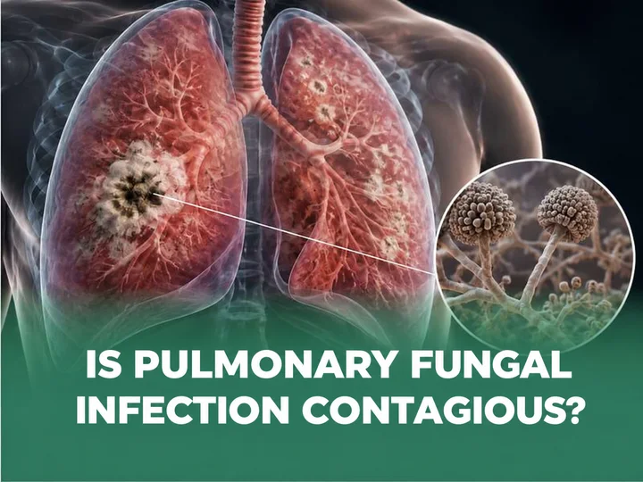



Pulmonary bullae are abnormal air filled spaces within the lung parenchyma. They may be associated with damage to lung tissue, long term smoking or conditions such as chronic obstructive pulmonary disease (COPD). Many cases cause no symptoms and are discovered incidentally on a chest X-ray or CT scan. However, large or ruptured bullae can compress the surrounding lung tissue, cause pneumothorax and impair breathing, requiring appropriate medical evaluation and monitoring.
Pulmonary bullae at a glance
- Are pulmonary bullae dangerous? They are often benign but may cause pneumothorax, infection or coughing up blood, particularly when they are large or have thin walls.
- When is urgent medical attention needed? Seek immediate care for sudden chest pain, rapidly worsening shortness of breath, coughing up blood, or fever accompanied by a cough producing purulent sputum.
- When may surgery be required? Surgery may be considered for giant bullae, progressively worsening shortness of breath, recurrent or persistent pneumothorax following rupture, recurrent infection, or coughing up blood.
What are pulmonary bullae?
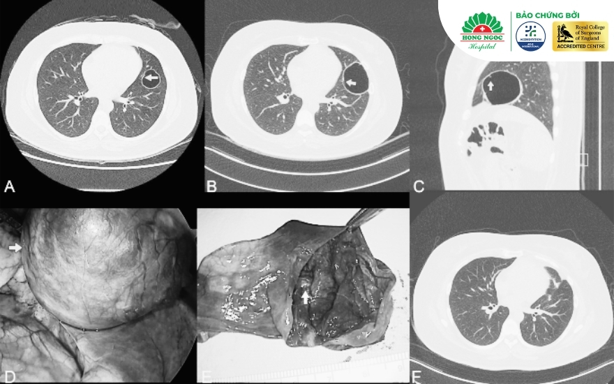
Pulmonary bullae, also referred to as bullous lung disease, develop when the walls between adjacent alveoli become thin or are destroyed, causing small air spaces to merge into one or more larger air filled spaces, typically exceeding 1 cm in diameter.
These bullae do not contribute to gas exchange. When they become very large, they may compress the surrounding healthy lung tissue and reduce the volume of functional lung available for breathing.
Causes of pulmonary bullae
The causes of pulmonary bullae are generally divided into two main groups:
Primary causes
Spontaneous bullae in young people: These are more common in tall, thin males aged 15–30. Rapid growth and greater negative pressure at the lung apices may contribute to stretching and disruption of the subpleural alveolar walls.
Genetic disorders and developmental abnormalities:
- Alpha-1 antitrypsin deficiency (AATD): Variants in the SERPINA1 gene reduce levels of a protein that protects lung tissue, leading to premature destruction of the alveoli.
- Birt-Hogg-Dubé syndrome (BHD): Variants in the FLCN gene cause pulmonary cysts, often in the lower lungs, and increase the risk of recurrent pneumothorax.
- Marfan syndrome and Ehlers-Danlos syndrome: Connective tissue disorders may reduce the structural strength of alveolar walls.
- Congenital structural abnormalities: These include congenital pulmonary airway malformation (CPAM) and bronchogenic cysts resulting from abnormal development of the fetal respiratory tract.
Secondary causes
Secondary bullae may develop through chronic inflammation or a one-way valve mechanism that traps air within the distal airways and alveoli:
- Smoking, vaping and air pollution: These exposures can trigger chronic inflammation and the release of enzymes that degrade collagen and elastin, damaging the alveolar walls.
- COPD and emphysema: Persistent air trapping and disease of the small airways can cause alveoli to enlarge and merge into bullae.
- Sequelae of severe infection: Pulmonary tuberculosis, lung abscess, necrotizing pneumonia caused by pathogens such as Staphylococcus aureus or fungi, and bronchiectasis may contribute to bulla formation.
- Fibrotic scarring with a check-valve mechanism: Air enters during inhalation but cannot escape effectively during exhalation, causing progressive overdistension of the alveoli.
- Previous trauma or surgery: Fibrotic scarring and tissue retraction may distort the small airways and cause localized air trapping.
Signs and symptoms of pulmonary bullae
During the early stages or when the bullae are small, the condition often causes no clear clinical symptoms. Pulmonary bullae are frequently discovered incidentally during a routine health examination or on a chest X-ray performed for another reason.
As the bullae enlarge or complications develop, patients may experience:
- Shortness of breath: The most common symptom. Breathlessness may gradually worsen during physical activity, brisk walking or climbing stairs as the bullae compress healthy lung tissue.
- Chest tightness or sudden chest pain: Patients may experience a dull ache or sharp pain on one side of the chest that worsens with deep breathing or coughing.
- Persistent cough: This may be dry or productive, particularly when bullae occur alongside chronic bronchitis.
- Fever and purulent or blood-stained sputum: If a lung infection is present, patients may develop fever, purulent sputum, coughing up blood and chest pain.
- Cyanosis, fatigue and rapid breathing: These may occur in advanced disease when blood oxygen levels fall significantly.
Medical advice: Anyone experiencing sudden chest pain accompanied by shortness of breath should seek immediate care at the nearest medical facility.

Classification of pulmonary bullae
According to Reid’s classical classification, as referenced in Shields’ General Thoracic Surgery and other medical literature, pulmonary bullae may be classified according to three main criteria: anatomical structure, size and degree of compression, and number of lesions.
By location and structural characteristics:
- Type I (superficial or subpleural bullae): These bullae have a narrow neck and lie immediately beneath the pleural surface, commonly at the lung apex. Their walls are extremely thin and prone to rupture, potentially causing spontaneous pneumothorax.
- Type II (superficial parenchymal bullae): These bullae are located in the peripheral lung parenchyma, have a broader base and communicate directly with the small airways.
- Type III (deep parenchymal bullae): These bullae are located deep within the lung parenchyma. They generally have thicker, more fibrotic walls and compress the surrounding lung tissue from within.
By distribution and degree of compression:
- Multiple bullae: Numerous bullae are scattered throughout one or both lungs. This pattern is commonly observed in people with COPD or pulmonary fibrosis.
- Giant bullae: A bulla occupying more than one third of one side of the chest. Giant bullae can severely compress the surrounding healthy lung tissue and often require surgical intervention.
By number of lesions:
- Solitary bulla: A single bulla develops within the lung parenchyma while the surrounding lung tissue remains normal. It usually causes no symptoms and is often discovered incidentally during a routine health examination or chest CT scan.
- Multiple bullae: Bullae of varying sizes develop in one or both lungs and may cause persistent shortness of breath, chronic cough and chest tightness.
Are pulmonary bullae dangerous? Common complications
Pulmonary bullae are noncancerous lesions. However, without appropriate monitoring and management, they may enlarge and lead to potentially life-threatening complications:
- Spontaneous pneumothorax: This is the most common and potentially serious complication. A thin-walled bulla may rupture when intrathoracic pressure increases, such as during forceful coughing or physical exertion. Air then enters the pleural space, potentially causing lung collapse and acute respiratory failure.
- Infection of a pulmonary bulla: Bacteria may infect a fluid-containing bulla, resulting in an abscess that can cause high fever, foul-smelling purulent sputum and severe chest pain.
- Coughing up blood: Damage to the bulla wall or surrounding blood vessels due to infection may cause anything from mild blood-streaked sputum to massive hemoptysis.
- Chronic respiratory failure: Prolonged compression by large bullae can significantly impair pulmonary ventilation and respiratory function.
Current treatment options for pulmonary bullae
Treatment aims to relieve compression of healthy lung tissue, prevent serious complications and improve respiratory function. The management plan depends on the size of the bullae, associated symptoms, underlying lung function and the presence of complications.
Medical management and monitoring
- Indications: Appropriate for small bullae that do not cause shortness of breath or other complications.
- Management options:
- Follow-up examinations every 6–12 months, accompanied by chest X-rays or CT scans when clinically indicated, to monitor changes in the bullae.
- Bronchodilators or preventive inhalers for patients with coexisting respiratory conditions such as COPD or asthma.
- Complete smoking cessation and avoidance of secondhand smoke.
Surgical intervention
Surgery may be considered to remove bullae causing symptoms or complications in appropriately selected patients.
- Indications for surgery:
- Giant bullae occupying more than 30–50% of one side of the chest.
- Progressive shortness of breath or significant compression of healthy lung tissue.
- Ruptured bullae causing recurrent or persistent pneumothorax.
- Recurrent infection or coughing up blood.
- Surgical approaches:
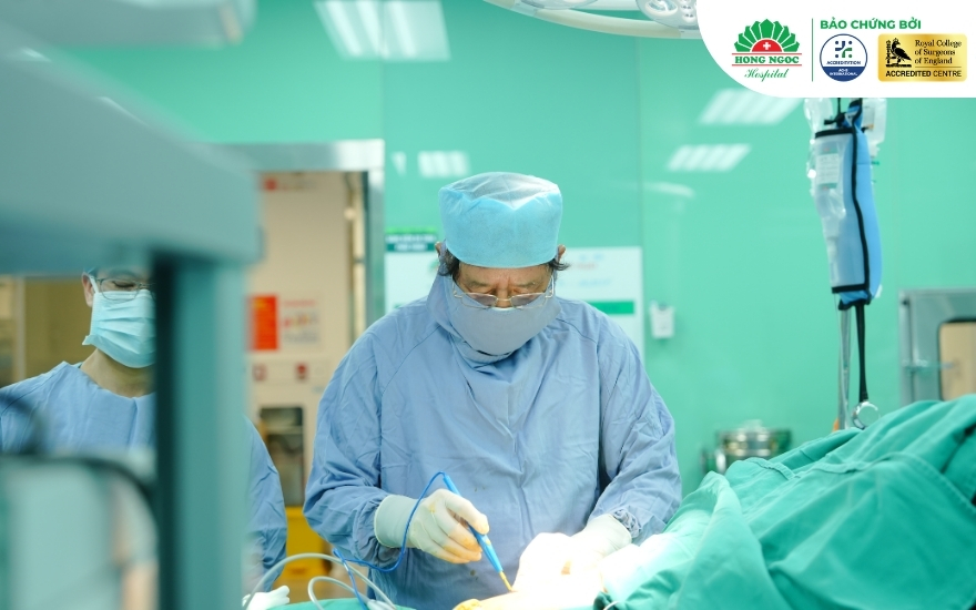
- Video-assisted thoracoscopic surgery (VATS): The standard minimally invasive approach in many suitable cases. The surgeon makes one to three small incisions, typically measuring approximately 1–2 cm, to insert a camera and surgical instruments, remove the bullae and repair the lung tissue. Potential benefits include less postoperative pain, smaller scars, faster recovery and a shorter hospital stay.
- Open thoracotomy: This approach may be required when there are extensive adhesions, very large bullae or severe complications that cannot be managed safely using thoracoscopic surgery.
Managing pulmonary bullae and preventing complications
To help slow the progression of bullous lung disease and support recovery after treatment, patients should follow these recommendations:
- Stop smoking completely: This is the most important measure for preventing further destruction of the alveolar walls.
- Limit exposure to dust, smoke and harmful chemicals: Wear appropriate protective masks when working in polluted environments.
- Seek prompt treatment for respiratory infections: Pneumonia and pulmonary tuberculosis should be properly managed to minimise further lung damage.
- Avoid sudden, strenuous physical activity: Do not lift excessively heavy objects or engage in activities that substantially increase intrathoracic pressure, such as scuba diving or heavy weight training, as these may increase the risk of bulla rupture. Patients should consult their doctor about appropriate levels of physical activity and activities involving major pressure changes, particularly if they have previously experienced a pneumothorax.
- Practise breathing exercises and pulmonary rehabilitation: Perform diaphragmatic breathing and pursed-lip breathing exercises under the guidance of a healthcare professional to improve breathing efficiency and respiratory function.
- Keep vaccinations up to date: Annual influenza vaccination and pneumococcal vaccination may help reduce the risk of respiratory infections.
- Maintain a balanced diet: Consume adequate protein, vitamins and other essential nutrients to support the immune system and overall health.
- Seek immediate medical attention for unusual symptoms: Sudden chest pain or rapidly worsening shortness of breath requires urgent medical assessment.
Diagnosis and treatment of pulmonary bullae at the Department of Thoracic Surgery, Hong Ngoc General Hospital
Early detection and appropriate treatment play a key role in managing pulmonary bullae and reducing the risk of serious complications such as pneumothorax. At Hong Ngoc General Hospital, patients have access to:
- Advanced diagnostic equipment: Digital X-ray and multidetector CT systems support the detection of pulmonary bullae, including small lesions, and help assess the extent of lung parenchymal damage.
- An experienced medical team: Leading specialists directly examine patients and develop individualised treatment plans based on disease severity, ranging from medical monitoring to thoracic surgery when indicated.
- International recognition for healthcare quality and surgical training: Hong Ngoc General Hospital:
- Holds ACHS International accreditation for healthcare standards.
- Is recognised by the Royal College of Surgeons of England as an international-standard surgical training centre.
If you or a family member has symptoms suggestive of pulmonary bullae or is seeking an appropriate minimally invasive surgical treatment, contact the Department of Thoracic Surgery at Hong Ngoc General Hospital via 0949 646 556 to arrange a consultation with a specialist.
Frequently asked questions
The following answers address common questions about the diagnosis and treatment of pulmonary bullae.
Are pulmonary bullae cancerous?
Pulmonary bullae are benign structural abnormalities of the alveoli and are not cancerous. However, bullae may obscure other abnormalities on imaging or coexist with conditions such as lung cancer or COPD. A specialist assessment may therefore be required to establish a differential diagnosis.
Can pulmonary bullae resolve on their own?
Once formed, pulmonary bullae generally do not disappear with medication. Medicines may help relieve breathlessness or treat associated infections, but they do not remove the bullae. When complete removal is clinically necessary, surgery may be considered.
Are pulmonary bullae hereditary?
Most pulmonary bullae are not directly inherited. However, a small proportion of cases may be associated with inherited disorders, such as alpha-1 antitrypsin deficiency.
How long can someone with pulmonary bullae live?
The prognosis depends on the underlying cause, extent of lung damage, respiratory function, coexisting lung disease and whether complications develop:
- If the bullae are small, remain stable and cause no complications, patients may live normally without any reduction in life expectancy. When surgery is indicated and successfully performed, respiratory function may improve considerably.
- In older patients with pulmonary bullae associated with severe COPD, the prognosis largely depends on the severity of the underlying respiratory impairment.
Do pulmonary bullae require surgery?
Not all patients with pulmonary bullae require surgery. The decision depends on the size of the bullae, the patient’s symptoms and the risk of complications. A specialist may recommend surgery when the bullae are large, cause significant breathlessness or have led to complications such as pneumothorax or recurrent infection.
Conclusion
Pulmonary bullae are benign structural abnormalities of the lung parenchyma, but they may lead to serious complications such as spontaneous pneumothorax, mediastinal compression and recurrent infection. They are commonly detected on a chest X-ray or CT scan performed when clinically indicated. Monitoring and treatment should be tailored to the characteristics of the lesions, the patient’s symptoms and any underlying lung disease.
—--------------------------------------------------------------------------
DEPARTMENT OF THORACIC SURGERY - HONG NGOC GENERAL HOSPITAL
- ACHS International accredited for healthcare standards
- Recognised by the Royal College of Surgeons of England as an international-standard surgical training centre
Addresses:
- Hong Ngoc Phuc Truong Minh General Hospital: 8 Chau Van Liem Street, Tu Liem Ward, Hanoi
- Hong Ngoc General Hospital – Yen Ninh: 55 Yen Ninh Street, Ba Dinh Ward, Hanoi
Hotline: 0949 646 556
Medical disclaimer: The information in this article is provided for general reference only and is not a substitute for diagnosis or treatment recommendations from a qualified medical specialist. Patients should attend a healthcare facility for an in-person examination and individualised medical advice.