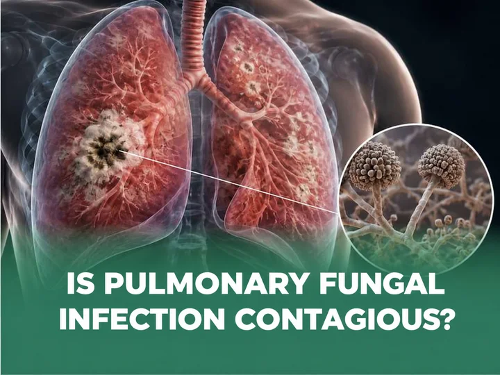



Foreign body airway obstruction is a dangerous emergency that can occur in both children and adults. If not recognized and managed promptly, it can become life-threatening within minutes. Equipping yourself with the knowledge to spot the warning signs early and to deliver first aid correctly is therefore the “key” to protecting your own life and the lives of those around you.
What is foreign body airway obstruction?
Foreign body airway obstruction occurs when food or another foreign object enters the larynx, trachea or bronchi instead of passing down the oesophagus. Once lodged, the object causes partial or complete blockage of the airway, impeding ventilation and leading to acute cerebral hypoxia. If not treated promptly, it can quickly prove fatal.
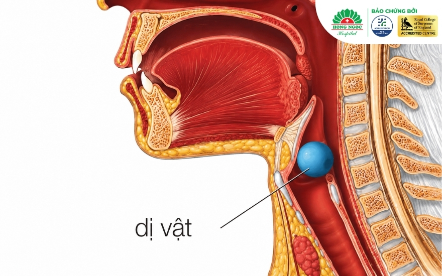
Common sites of airway foreign body lodgement
Depending on the size and shape of the object and the victim’s position at the time of choking, a foreign body may lodge at different sites:
Laryngeal foreign body
Location and risks: Located at the uppermost part of the respiratory tract (at the epiglottis and the glottis), this is a particularly dangerous site for a foreign body to lodge. It can readily cause acute asphyxia and carries a high risk of death if not managed promptly.
Clinical presentation:
- A foreign body lodged here can rapidly compress and obstruct the airway.
- A typical penetration syndrome develops: violent coughing and choking, cyanosis, sudden breathing difficulty, hoarseness or complete loss of voice.
Tracheal foreign body
Location and risks: The foreign body lodges in the segment of the airway that extends from just below the larynx to the carina, where the trachea divides into the two main bronchi. This is an extremely urgent emergency, because a foreign body in the trachea can move freely and may completely occlude the airway.
Clinical presentation:
- A tracheal foreign body is typically mobile: as the patient inhales and exhales or coughs, the airflow pushes it up and down.
- On auscultation, or when the ear is placed close to the patient’s mouth, a “flapping flag” sound (audible slap) or a wheezing or stridulous sound may be heard as the object strikes the tracheal wall.
Bronchial foreign body
Location: The right main bronchus is usually shorter, wider and more vertical than the left. As a result, most bronchial foreign bodies lodge in the right main bronchus or the right lower lobe bronchi. If left in place, a bronchial foreign body can lead to complications such as pneumonia, atelectasis, or collapse of a lung lobe due to obstruction.
Clinical presentation:
- After the initial episode of violent coughing and choking, the foreign body remains fixed in one bronchus.
- Breath sounds are reduced or absent over the obstructed lung.
Causes of foreign body airway obstruction
In practice, foreign body airway obstruction can occur at any age, but it is most common in children and older people:
In children:
- Young children tend to put small objects (marbles, buttons, toys, melon seeds, sunflower seeds) into their nose or mouth and then accidentally inhale them into the airway.
- Children’s molars are not yet fully developed, so they cannot chew food thoroughly (they are prone to choking on beans, chestnuts, jelly and smooth, round fruits such as longan and rambutan).
- Children often eat while laughing, crying, running or jumping around.
In adults and older people:
- Talking while eating, or eating too quickly.
- Impaired swallowing reflex due to ageing or neurological damage (for example, after a stroke or in Parkinson’s disease).
- Holding small items in the mouth while working (toothpicks, hairpins and the like); choking on fish bones, chicken bones, or fragments of cracked dentures that fall into the airway.
- Dentures that become dislodged during eating or while dozing off and slip down into the larynx, trachea or bronchi.
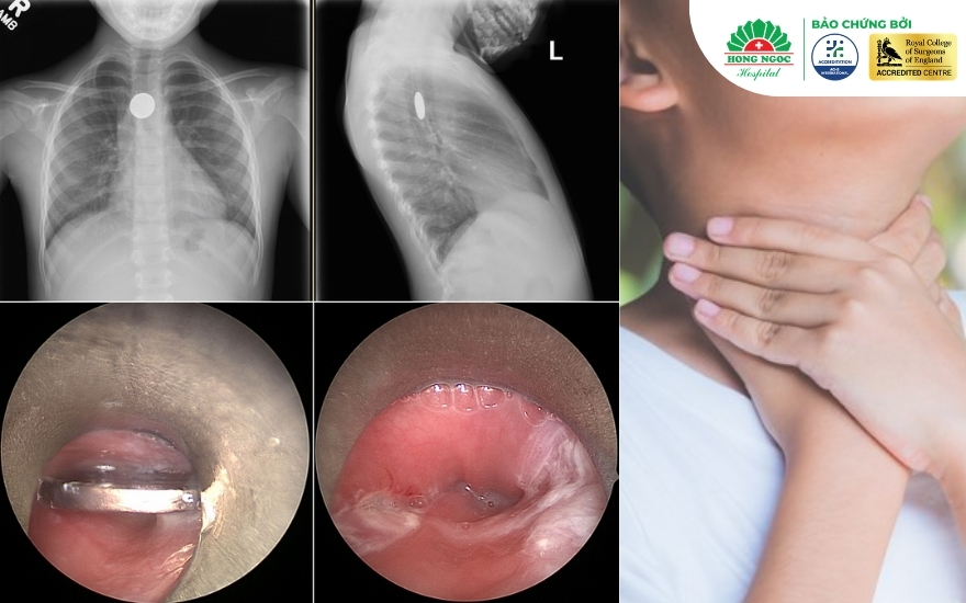
Signs of foreign body airway obstruction
When a foreign object (food, a toy or another item) enters the airway, the body reacts differently depending on the degree of obstruction. Correctly identifying its severity enables bystanders to respond promptly and appropriately.
Partial airway obstruction
At this level, air can still pass around the object, and the victim retains the body’s protective reflexes.
- Strong, clear cough: The victim coughs continuously and loudly. This is a good reflex that helps expel the foreign body.
- Still able to make sounds: The person has only just begun choking but can still speak, cry or answer when asked.
- Noisy breathing: Wheezing or mild stridor may be heard when the victim breathes in.
- Little change in appearance: The victim is alert, and the skin may look slightly flushed from the effort of coughing, but there is no cyanosis.
Complete airway obstruction
This is a life-threatening condition because airflow is completely or almost completely blocked.
- Unable to speak or cry: The foreign body has blocked all or nearly all of the airway.
- Weak or absent cough: The cough is feeble and ineffectual, or makes no sound at all, because there is not enough airflow to generate a cough.
- Characteristic sign (the universal choking sign): The victim clutches their throat with both hands and shows obvious panic and fear.
- Silent, open-mouthed struggle: The victim opens their mouth but makes no sound, or produces only a very faint high-pitched wheeze.
- Severe respiratory distress: The chest heaves in an effort to breathe, but no air enters, and the intercostal spaces are deeply retracted.
- Cyanosis: The lips, fingernails and face turn bluish because of severe oxygen deprivation.
- Loss of consciousness: The victim quickly collapses, becomes limp and stops breathing unless the airway is cleared immediately.
Penetration syndrome
This is the body’s violent protective reflex when a foreign object touches the mucosa of the larynx and trachea:
- A patient who is eating or playing suddenly develops violent coughing, choking and paroxysmal asphyxia.
- The face turns intensely red, then rapidly becomes cyanotic around the lips and fingertips.
- Wheezing and stridor are present.
- Some victims may clutch their throat, the commonly recognized sign of choking. However, this sign does not appear in every case.
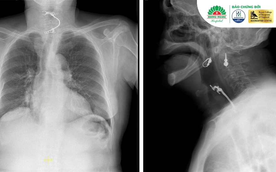
Dangerous complications if not managed promptly
If an airway foreign body is not managed promptly, the patient faces a range of severe acute and chronic complications:
- Acute death: Caused by acute asphyxia and extensive cerebral hypoxia, leading to cardiac arrest.
- Atelectasis: A foreign body that completely blocks a bronchus prevents air from reaching the corresponding lung lobe.
- Obstructive emphysema: The foreign body acts as a one-way valve, allowing air in but not out. This overinflates the alveoli, which may rupture and lead to pneumothorax or pneumomediastinum.
- Recurrent pneumonia and lung abscess: An organic foreign body (such as a seed or food) retained in the bronchus for a long time provokes inflammation and infection, producing purulent secretions and destroying lung tissue.
- Post-obstructive bronchiectasis: Prolonged chronic infection destroys the fibromuscular structure of the bronchial wall.
Instructions for managing and administering first aid for foreign body airway obstruction
When someone is found to be choking on a foreign body, correct first aid before the patient reaches a medical facility is critical to survival.
Initial principles
- Alert and able to cough: Encourage the patient to keep coughing forcefully to expel the foreign body. Do not blindly sweep the throat with your fingers, as this may push the object deeper into the trachea (or bronchus).
- Choking, unable to speak or cough, cyanotic: Begin emergency first-aid manoeuvres immediately.
Emergency first-aid techniques
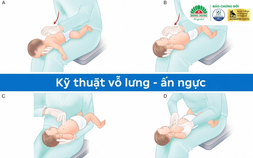
A. Infants under 1 year (back blows and chest thrusts)
According to the Pediatric Basic Life Support guidelines of the American Heart Association (AHA), the first-aid technique for an infant under 1 year old who is choking on a foreign body is as follows:
- Back blows (5): Lay the infant face down along your forearm, with the head lower than the chest, firmly supporting the jaw. Using the heel of your other hand, deliver five firm, sharp blows between the infant’s shoulder blades.
- Chest thrusts (5): Turn the infant face up along your other forearm, keeping the head lower than the trunk. Using two fingers (index and middle), give five firm thrusts to the lower half of the breastbone.
- Alternate the two steps until the infant cries or the object is expelled. Do not perform abdominal thrusts (the Heimlich manoeuvre) on infants under 1 year.
If the infant loses consciousness:
- Call for emergency help immediately: Place the infant on a firm, flat surface and call 115 or your local emergency number.
- Check the airway: Open the infant’s mouth. If the foreign body is clearly visible, remove it with your fingers. Never sweep the throat blindly or try to feel for the object with your fingers, as this may push it deeper.
- Begin cardiopulmonary resuscitation (CPR):
- Chest compressions: Using two fingers (or two thumbs if encircling the chest), press hard and fast on the lower half of the breastbone at a rate of 100 to 120 compressions per minute, to a depth of about one-third of the chest, or roughly 4 cm.
- Rescue breaths: After 30 compressions, give two rescue breaths, covering the infant’s mouth and nose with your mouth to form a tight seal.
- Repeat: Continue cycles of 30 compressions and two breaths until medical personnel arrive or the infant responds. Each time you open the airway to give breaths, check whether the object has moved into the mouth and remove it if so.
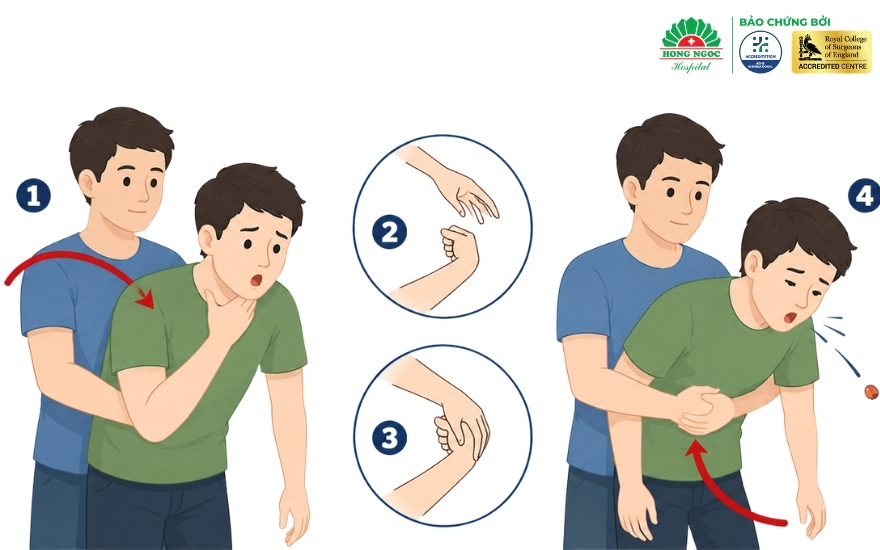
B. Children over 1 year and adults (the Heimlich manoeuvre)
According to the American Heart Association (AHA) guidelines on cardiopulmonary resuscitation and first aid for airway foreign bodies, the emergency first-aid procedure for a choking child over 1 year old is as follows:
If the victim is conscious:
- Stand behind the victim and wrap your arms around their waist.
- Make a fist and place the thumb side of the fist (the bony knuckle of the thumb) against the upper abdomen, just above the navel and well below the breastbone (avoid pressing on the sternum or xiphoid process).
- Grasp the fist with your other hand and deliver a firm, sharp thrust inward and upward (in a J-shaped motion). Give five thrusts in succession, and repeat until the object is expelled and the victim can cough or breathe again.
If the victim is unconscious: Lay the person on their back and kneel beside them. Place the heel of your hand on the upper abdomen and press hard upward. Begin cardiopulmonary resuscitation (CPR) and call 115 or your local emergency number immediately.
Note: The Heimlich manoeuvre may not be suitable in certain cases, such as late pregnancy or people with special physical conditions. Because of the complexity of these cases, take the patient to the nearest medical facility without delay, or call the emergency number 115 so that medical personnel can provide timely assistance.
How are airway foreign bodies diagnosed and removed?
Diagnosing and removing an airway foreign body is a rigorous medical process that combines initial history-taking, clinical examination, and modern imaging and interventional methods.
1. Taking the history (medical history and the choking episode)
- Medical history: The doctor gathers detailed information about the suspected foreign body (a seed, plastic toy, bone, metal object or other item), when the incident occurred and the initial symptoms.
- Identifying penetration syndrome: The doctor asks whether the patient recently experienced a sudden fit of violent coughing, cyanosis, breathing difficulty or panic. This is the most important clue in diagnosis.
2. Clinical respiratory examination
- The doctor listens to the lungs with a stethoscope to assess airflow.
- The doctor looks for abnormal signs such as tracheal stridor, reduced or absent breath sounds on one side (when the foreign body obstructs a bronchus), or localized wheezing.
3. Imaging (X-ray and CT scan)
- Chest X-ray (frontal and lateral): Quickly detects radiopaque foreign bodies (metal, bone and the like). For radiolucent objects (grapefruit seeds, plastic items), the X-ray shows indirect signs such as atelectasis, obstructive emphysema or pneumothorax.
- Chest computed tomography (CT) scan: Indicated in complex cases, where a deeply lodged or long-retained foreign body is suspected, to determine its exact location, size and the surrounding tissue damage.
4. Airway endoscopy
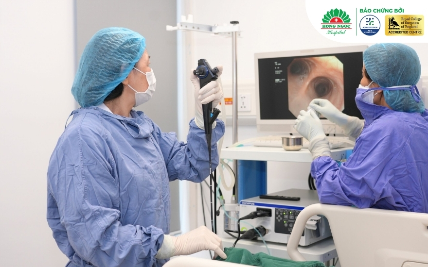
- Diagnostic endoscopy: When there is clinical suspicion but the X-ray findings are unclear, the doctor performs bronchoscopy (flexible or rigid) to directly inspect the entire airway, from the larynx and trachea to the bronchial branches.
- Endoscopic removal:
- Once the foreign body is found, the doctor passes specialised instruments (forceps, snare baskets, balloon catheters) through the bronchoscope to grasp and extract it safely.
- The procedure is always performed with airway control and appropriate anaesthesia or sedation to ensure maximum patient safety.
Preventing choking
Taking proactive steps to prevent airway foreign body obstruction is the best way to protect yourself and your family from this dangerous accident:
- For young children:
- Keep small items out of children’s reach: beads, button batteries, small detachable toy parts, pen caps, screws and the like.
- Do not give children nuts and seeds (sunflower seeds, chestnuts, peanuts), hard jelly cubes or fruit with the pits or seeds still in, until they have developed adequate chewing skills.
- Do not feed children or give them drinks while they are crying or laughing.
- Teach children to sit still while eating, and not to run, jump or play while there is food in their mouths.
- For adults:
- Chew thoroughly and swallow slowly; avoid talking or laughing excessively while eating.
- Remove bones carefully when preparing meat and fish for children and older people.
- Have dentures checked regularly to make sure they fit securely. Do not hold small objects in the mouth while working.
- For older people and people with neurological conditions: Prepare soft food, cut it into small pieces or puree it to avoid swallowing difficulties.
Managing airway foreign bodies at Hong Ngoc General Hospital
At Hong Ngoc General Hospital, airway foreign bodies are managed according to a standardised clinical protocol designed to ensure accuracy, safety and the best possible care for each patient group:
1. Imaging assessment and accurate diagnosis
- As soon as a patient with a suspected foreign body is admitted, doctors carry out an urgent clinical examination.
- Modern imaging techniques (digital X-ray and multislice chest CT) are performed rapidly to determine the exact location, size and shape of the foreign body, as well as the extent of airway injury.
2. Interventional airway endoscopy (foreign body removal)
- Flexible or rigid bronchoscopy systems allow direct visualisation of the entire bronchial tree.
- Doctors remove the foreign body gently and precisely using specialised instruments (forceps, snares and balloon catheters), minimising invasiveness and trauma to the mucosa.
3. Comprehensive multidisciplinary coordination
- Anaesthesia and critical care: Ensures safe airway control, keeping the patient pain-free and free from coughing or panic throughout the procedure.
- Ear, nose and throat (ENT) and paediatrics: Work directly together on assessment and specialist management for young children, or when the foreign body is located in the pharynx or larynx.
- Thoracic surgery: Stands ready to provide consultation and surgical intervention in complex cases, such as a foreign body embedded deep in the bronchial wall, or when serious complications have developed.
4. Management of complications and post-procedure care
- Doctors irrigate the airway and suction out pus and infected material, or treat mucosal damage caused by a long-retained foreign body.
- Respiratory function is closely monitored, anti-inflammatory treatment and antibiotics are given if infection is present, and recovery is assessed before the patient is discharged.
Frequently asked questions about foreign body airway obstruction
What should I do if someone is choking but can still breathe?
This is a case of partial airway obstruction. At this stage, the cough reflex is the body’s most effective natural defence. You should not perform abdominal thrusts or back blows straight away, and you must never give the person water to drink.
Stay calm and encourage the person to keep coughing forcefully to expel the foreign body on their own. If the object has not come out after a few minutes of coughing, or if signs of breathing difficulty or wheezing appear, take the person to the nearest medical facility immediately.
Should you sweep the throat with your fingers when someone is choking?
No. A blind finger sweep (when the object cannot be seen) carries a very high risk of pushing the foreign body deeper into the airway. Use your fingers to remove the object only when it has moved into the mouth or throat and is clearly visible.
What should I do if a child is choking?
The first step is to assess the child’s breathing:
If the child is still pink, and can cry or cough: Let the child keep coughing, reassure them and take them to hospital promptly.
If the child cannot cry, is turning blue or has a weak cough: Perform emergency first aid immediately: back blows and chest thrusts (for infants under 1 year) or the Heimlich manoeuvre (for children over 1 year), while asking a bystander to call the emergency number 115.
Can the Heimlich manoeuvre be used on infants under 1 year?
No. Infants under 1 year have a relatively large liver that sits low in the abdomen, and their abdominal organs are very soft and delicate. Abdominal thrusts (the Heimlich manoeuvre) at this age can cause serious injuries such as liver rupture and internal bleeding. For infants under 1 year, the only medically recommended first-aid method is back blows and chest thrusts.
Is it a problem if I keep coughing after the object is gone?
A mild cough after the foreign body has come out may be caused by scratching, irritation or mild inflammation of the airway lining. However, if the cough persists, produces phlegm or pus, or is accompanied by fever, breathing difficulty or chest pain, see a doctor immediately. These may be signs that part of the object (fragments) remains in the airway, or that complications such as pneumonia have developed.
Can an airway foreign body come out on its own?
A foreign body can be expelled by a strong cough reflex, but this is uncommon and occurs only with small, smooth objects. Most airway foreign bodies become lodged because of reflex spasm of the airway muscles. For this reason, you should not wait for the object to “come out by itself”, and should seek medical care promptly. A foreign body left in place can lead to infection, abscess or lung collapse.
Can an X-ray detect an airway foreign body?
An X-ray directly detects only radiopaque objects (such as metal fragments, screws, dentures and thick animal bones). Radiolucent objects (such as seeds, plastic toys and food) are not directly visible on X-ray, but the image helps doctors identify indirect signs such as atelectasis or obstructive emphysema. In these cases, a CT scan or bronchoscopy allows a more accurate diagnosis.
When is bronchoscopy needed to remove a foreign body?
Bronchoscopy is the “gold standard” and is indicated when:
The patient has symptoms suggesting a foreign body (a choking episode, a persistent cough, or wheezing in one lung) even though no foreign body is seen on X-ray.
A foreign body lodged in the airway has been confirmed on imaging.
The object needs to be removed safely and pus and infected material caused by a long-retained foreign body need to be cleared.
Conclusion
An airway foreign body is a medical emergency that can be directly life-threatening within a matter of minutes. Recognising the signs early and applying correct first-aid techniques within the “golden time” are the key to saving the victim’s life. Remember that even after the foreign body has been removed, the patient still needs to be examined by a doctor at a medical facility for a full assessment of any damage to the respiratory tract.
—--------------------------------------------------------------------------
THORACIC SURGERY DEPARTMENT, HONG NGOC GENERAL HOSPITAL
- Accredited by ACHS International for healthcare standards
- Certified as a surgical training centre under the standards of the Royal College of Surgeons (RCS)
Addresses:
- Hong Ngoc General Hospital, Phuc Truong Minh campus: No. 8 Chau Van Liem Street, Tu Liem Ward, Hanoi
- Hong Ngoc General Hospital: No. 55 Yen Ninh Street, Ba Dinh Ward, Hanoi
Hotline: 0949 646 556
Note: The information in this article is for reference and educational purposes only and is in no way a substitute for medical diagnosis, advice or treatment. If someone shows signs of airway foreign body obstruction, first aid should be given only by a person who is well versed in the relevant knowledge and techniques. At the same time, call the emergency number 115 or go to the nearest medical facility without delay so that the patient can be treated promptly.