Lumbar spondylolisthesis is a condition characterized by the displacement of a vertebra from its normal anatomical position, leading to pain and discomfort that can significantly affect daily activities.
In this article, Hong Ngoc General Hospital outlines the causes, clinical manifestations and treatment options for lumbar spondylolisthesis.
What is lumbar spondylolisthesis?
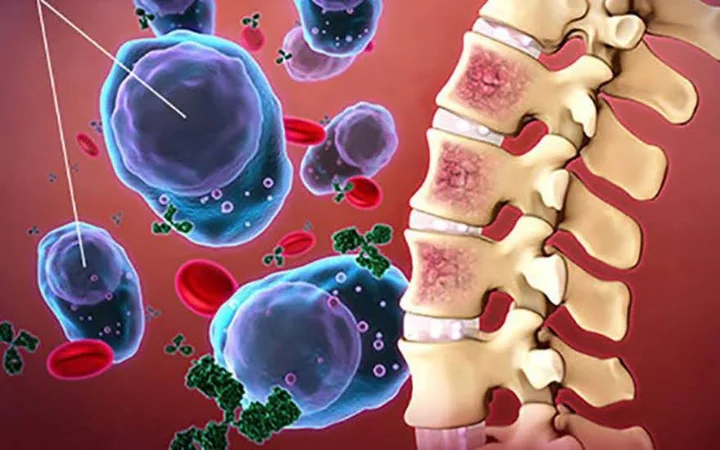
Lumbar spondylolisthesis is a condition in which a vertebral body slips out of its normal anatomical alignment, either anteriorly or posteriorly relative to the adjacent vertebra below. Patients with lumbar spondylolisthesis are often burdened by persistent lower back pain radiating to one or both lower extremities, accompanied by impaired mobility and difficulty with daily activities.
If left untreated, lumbar spondylolisthesis may lead to serious complications, including limb numbness, motor dysfunction, muscle atrophy, and in severe cases, permanent disability.

Causes of lumbar spondylolisthesis
Congenital lumbar spondylolisthesis: This form results from developmental abnormalities during adolescence, including hypoplasia of the articular processes and abnormal orientation of the facet joints, which may be directed posteriorly (often associated with spina bifida) or medially.
Isthmic spondylolisthesis (pars interarticularis defect): This type is associated with lesions of the pars interarticularis, such as defects or stress fractures involving the segment connecting adjacent lumbar vertebrae, most commonly at L5–S1. Etiologies include repetitive stress fractures, elongation of the pars due to continuous cycles of fracture and healing, or acute fractures of the pars region. This differs from traumatic spondylolisthesis occurring outside the pars interarticularis.
Degenerative lumbar spondylolisthesis: Degenerative changes of the spine, particularly intervertebral disc degeneration and facet joint arthropathy, lead to spinal instability and vertebral slippage. This type most commonly occurs at the L4–L5 level.
Pathological lumbar spondylolisthesis: Certain conditions, such as spinal infections or malignancies, can compromise the structural integrity of the vertebrae, resulting in vertebral displacement.
Traumatic lumbar spondylolisthesis: In some cases, acute trauma causing fractures of the pedicles or facet joints can destabilize the spine and lead to vertebral slippage.
Iatrogenic (post-surgical) spondylolisthesis: Spinal procedures such as laminectomy or extensive decompression combined with facet joint resection may result in postoperative instability and subsequent spondylolisthesis, particularly in patients with pre-existing spinal instability.
Clinical manifestations of lumbar spondylolisthesis
Although lumbar spondylolisthesis is not typically life-threatening, it can significantly impair a patient’s quality of life. The condition presents with a range of clinical symptoms; however, in the early stages, manifestations such as mild lower back pain or intermittent leg numbness may be subtle and nonspecific, leading many patients to delay medical evaluation.
Early-stage (lumbar pain phase): Pain is usually progressive in nature. Initially, patients may experience discomfort during activities such as walking, bending, extension, or prolonged standing. Over time, pain intensifies with movement and positional changes, particularly when transitioning from sitting to standing. Patients may need to pause frequently while walking due to pain. As the condition progresses, pain may radiate to the buttocks, thighs, calves, and feet, often accompanied by paresthesia. Symptoms may be exacerbated by coughing or sneezing.
Advanced stage: Patients continue to experience persistent pain, often associated with lumbar muscle spasm and tightness of the inner thigh musculature. Postural abnormalities may develop, including a forward-flexed gait, loss of normal lumbar lordosis, scoliosis, or kyphotic deformity. Pain may occur in recurrent episodes, becoming more severe, frequent, and sometimes described as burning in nature. In more advanced cases, a palpable step-off deformity in the lumbar region (step sign) may be detected. Prolonged immobility can lead to muscle atrophy, and gait disturbances may resemble those seen in early childhood ambulation.

Diagnostic approaches for lumbar spondylolisthesis
Clinical evaluation
Lumbar spondylolisthesis can be clinically diagnosed based on characteristic findings associated with two main syndromes: the spinal syndrome and nerve root compression syndrome.
Spinal syndrome: Patients typically present with lower back pain, lumbar muscle spasm, and tightness of the inner thigh muscles, leading to difficulty in ambulation and restricted mobility. A palpable step-off deformity in the lumbar region may be detected. Pain is often exacerbated by axial loading and positional changes. In advanced cases, patients may develop noticeable postural and gait abnormalities.
Radicular (nerve root compression) syndrome: Pain may radiate to the buttocks, thighs, calves, and feet, often worsening with coughing or sneezing. Additional manifestations include sensory disturbances, reduced mobility, muscle atrophy, and in severe cases, motor weakness or paralysis. Some patients may experience persistent pain even at rest.
Paraclinical investigations
- Plain radiography (X-ray): Conventional X-rays are essential for determining the presence, location, and degree of vertebral slippage.
- Computed tomography (CT scan): CT imaging provides detailed visualization of bony structures, allowing accurate assessment of the degree of slippage, as well as associated lesions involving the pars interarticularis, facet joints, and potential spinal canal stenosis. It is particularly useful in cases of isthmic defects.
- Magnetic resonance imaging (MRI): MRI is indicated to identify the underlying causes of nerve root compression and to evaluate soft tissue structures, including intervertebral discs and neural elements, thereby assessing the extent of neural compression in lumbar spondylolisthesis.

Treatment options for lumbar spondylolisthesis
Most patients with lumbar spondylolisthesis can be managed conservatively with medical therapy, including pharmacological treatment, physical therapy, bracing, activity modification, and adequate rest.
However, in severe cases or when serious complications arise, surgical intervention such as spinal fusion may be indicated.
Conservative management
- Pharmacological therapy: Medications commonly include analgesics and anti-inflammatory agents, neuropathic pain modulators and antidepressants, muscle relaxants, neurotrophic agents, as well as gastric acid–reducing drugs for gastrointestinal protection. Dosage and administration must strictly follow medical prescription, as prolonged use of analgesics may adversely affect the gastrointestinal system and hepatic function.
- Physical therapy and rehabilitation: Structured rehabilitation programs incorporating gradual stretching and strengthening exercises help reduce mechanical stress on the lumbar spine. These exercises aim to enhance the strength of the paraspinal, abdominal, and thigh muscles, thereby alleviating pain and improving functional capacity. Rehabilitation should be performed under the supervision of qualified physiatrists or rehabilitation specialists.
In addition, patients are advised to modify daily activities, rest during acute pain episodes, and pursue weight reduction in cases of obesity to decrease spinal load.
Surgical management
Surgery is considered a last-line treatment for patients with severe lumbar spondylolisthesis who experience persistent pain unresponsive to conservative therapy after 6–12 months, or who develop complications such as muscle atrophy, motor deficits in one or both lower limbs, or bladder dysfunction.
Surgical techniques typically include vertebral reduction, spinal stabilization using pedicle screw fixation, and posterior interbody fusion. The primary objectives of surgery are neural decompression, elimination of abnormal vertebral motion, restoration of spinal stability, and promotion of solid bony fusion. Pain is expected to resolve once stable fusion is achieved.
Reader may also be interested in:
- Stages of disc herniation: recognizing each stage for appropriate management
- Lumbar spondylolisthesis: causes, symptoms and treatment options
However, surgical intervention carries inherent risks, including postoperative pain and surgical site infection. Outcomes depend on multiple factors, particularly the patient’s overall health status and disease severity. In patients with concomitant osteoporosis, surgical failure may occur due to poor screw fixation and suboptimal fusion rates. Early intervention prior to the onset of complications such as muscle atrophy, urinary retention, or paralysis generally yields better outcomes. Higher-grade vertebral slippage is associated with increased surgical complexity and a higher risk of complications.
Prognosis and recommendations
Treatment outcomes for lumbar spondylolisthesis depend on several factors, with early and accurate diagnosis being the most critical for establishing an appropriate management plan. Patients presenting with suggestive symptoms should seek prompt evaluation by musculoskeletal specialists. Self-medication without medical supervision is strongly discouraged, as it may lead to disease progression and poorer clinical outcomes.

With extensive experience in the management of musculoskeletal disorders, the Department of Orthopedics and Musculoskeletal Medicine at Hong Ngoc General Hospital is a trusted destination in Hanoi for the safe and effective treatment of lumbar spondylolisthesis.
- Highly qualified and experienced medical team: Dr. Pham Van Cuong, PhD, with over 35 years of experience, has successfully performed numerous complex hip and knee replacement surgeries as well as orthopedic trauma procedures. Dr. Nguyen Thi Ngoc, PhD, brings more than 30 years of expertise in conservative management of musculoskeletal conditions, with a focus on minimizing the impact on patients’ underlying comorbidities. The department is further supported by a team of skilled physicians, including MSc. Dr. Ngo Thi Trang, MSc. Dr. Hoang Thi Phuong Thao, MSc. Dr. Phan Thi Sinh, among others.
- Advanced medical equipment and technology: The hospital is equipped with state-of-the-art systems imported from the United States, ensuring optimal diagnostic accuracy and treatment outcomes. The SIGNA Prime MRI system, integrated with AIR™ reconstruction technology, enables early detection of tumors and musculoskeletal lesions. The 128-slice CT scanner reduces radiation exposure by up to 82% while delivering high-resolution imaging of the cervical and lumbar spine in a shorter acquisition time. Additional advanced equipment includes digital X-ray systems, Hologic bone densitometry (DXA) compliant with WHO standards, and the Logiq P7 musculoskeletal ultrasound system.

In addition, the department adopts a multidisciplinary approach by integrating Physical Therapy and Rehabilitation into treatment protocols. Advanced therapeutic systems imported from Germany such as shockwave therapy, radiofrequency therapy, and magnetic field therapy are combined with specialized manual manipulation techniques from Austria, enabling patients to restore mobility more rapidly.
Patients receiving care at Hong Ngoc General Hospital also benefit from a wide range of premium amenities:
- A clean, modern hospital environment with complimentary Wi-Fi, on-site cafés, and dining facilities
- Personalized follow-up schedules with proactive appointment reminders for each patient
- Efficient direct billing procedures, ensuring optimal insurance coverage and patient convenience
- Weekend services (including Saturdays and Sundays) with no additional charges
Note: The information provided in this article by Hong Ngoc General Hospital is for reference purposes only and does not replace professional medical diagnosis or treatment. Patients are advised not to self-medicate. For an accurate assessment of their condition, individuals should visit a healthcare facility for direct examination, diagnosis, and appropriate treatment consultation by qualified physicians.
For more health-related information, please follow the official fanpage of Hong Ngoc General Hospital:https://www.facebook.com/BenhvienHongNgoc