Osteomyelitis is a serious pathological condition affecting bone tissue, most commonly caused by bacterial infection or traumatic injury. If not diagnosed and treated promptly, the disease may lead to severe complications such as bone necrosis, skeletal deformity, or permanent functional impairment. A thorough understanding of its etiology, clinical manifestations, and treatment strategies is essential for early intervention and prevention of adverse outcomes.
What is osteomyelitis?
Osteomyelitis is a serious infectious disease of bone tissue caused by pathogenic microorganisms, most commonly pyogenic bacteria, Mycobacterium tuberculosis, or fungi. The infection damages the bone architecture, leads to local vascular obstruction, and results in suppuration and bone necrosis. Without timely treatment, the infection may spread, causing severe complications and, in advanced cases, posing a life-threatening risk. Osteomyelitis occurs more frequently in males, with incidence increasing with age.
Osteomyelitis spreads through three main routes:
Route 1: Hematogenous spread (via the bloodstream)
When bacteria circulating in the bloodstream invade bone tissue, they trigger an acute inflammatory response, damaging the periosteum and spreading within the bone, ultimately leading to bone necrosis. In children, bones are softer and more vulnerable, making it easier for large subperiosteal abscesses to form along the bone surface. During this process, necrotic bone fragments (sequestra) may separate from healthy bone.
In the chronic stage, persistent inflammation results in the accumulation of inflammatory cells and cytokines, promoting progressive bone destruction and fibrous tissue proliferation. New bone formation may occur around the affected area, creating a structure known as involucrum. If an abscess ruptures, purulent material can extend into surrounding soft tissues, causing secondary infection and the formation of draining sinus tracts through the skin.
Route 2: Contiguous spread from adjacent infected tissues or open wounds
Spread from neighboring infected soft tissues or through open wounds accounts for approximately 80% of osteomyelitis cases. This form is typically polymicrobial and most commonly affects the foot. Osteomyelitis frequently develops in bones compromised by trauma, surgical procedures, or radiotherapy, as well as in areas adjacent to pressure ulcers, such as the hip region and the sacrum.
Route 3: Vascular and neuropathic disorders leading to osteomyelitis of the lower extremities
Conditions such as peripheral vascular disease and diabetic neuropathy are significant risk factors for osteomyelitis of the lower limbs. Impaired blood circulation reduces the delivery of oxygen and nutrients to bone tissue, rendering it more susceptible to injury and hindering recovery from minor trauma or infection.
Concurrently, sensory neuropathy leads to loss of protective sensation in the feet, preventing patients from recognizing ulcers, wounds, or abscesses. This delay in detection allows pathogenic microorganisms to invade and spread to the underlying bone.
Causes of osteomyelitis
Osteomyelitis may arise from multiple etiologies, with the primary causative agents including common pyogenic bacteria, Mycobacterium tuberculosis, and fungal pathogens, all of which carry a high risk of severe complications if not promptly and appropriately managed.
Common bacterial pathogens:
In adults, the most frequent causative organism of osteomyelitis is Staphylococcus aureus. Other identified pathogens include coagulase-negative staphylococci, Streptococcus species, gram-negative bacilli, and anaerobic bacteria.
In neonates, common pathogens include Staphylococcus aureus, Streptococcus agalactiae, and Escherichia coli, typically identified through blood or bone cultures. In children older than one year, osteomyelitis is most commonly caused by Staphylococcus aureus, Streptococcus pyogenes, and Haemophilus influenzae.

Mycobacterial infection:
Skeletal tuberculosis occurs when Mycobacterium tuberculosis spreads hematogenously to bone tissue. In rare cases, infection may originate from contiguous spread from adjacent tuberculous lymph nodes. In addition to Mycobacterium tuberculosis, non-tuberculous mycobacteria such as Mycobacterium marinum, Mycobacterium avium–intracellulare, Mycobacterium fortuitum, and Mycobacterium gordonae have also been reported as causative agents of bone infection.
Fungal pathogens:
Fungal osteomyelitis is more commonly observed in elderly individuals and immunocompromised patients. Reported causative fungi include Coccidioides species (coccidioidomycosis), Blastomyces species (blastomycosis), Cryptococcus species, and Sporothrix species (sporotrichosis).
Clinical manifestations of osteomyelitis
Osteomyelitis may present with a wide spectrum of clinical manifestations, depending on the severity, duration, and underlying etiology of the infection.
- Bone pain: The most common symptom, typically dull and persistent, often worsening at night. In acute bacterial infection, the pain may be severe and abrupt in onset.
- Localized swelling, warmth, and erythema over the affected bone: These signs are more evident when the infection involves superficial bones or is associated with soft tissue abscesses or cellulitis.
- Sinus tract formation: Draining sinus tracts are commonly observed in chronic osteomyelitis or skeletal tuberculosis. Purulent discharge from the sinus opening is a characteristic sign of long-standing infection.
- Soft tissue involvement and bone exposure: Seen in cases of open fractures or penetrating joint injuries, creating a direct communication between bone tissue and the joint space.
- Distal limb necrosis: Ischemic changes leading to dry gangrene of the distal extremities may occur due to compromised blood supply, particularly in patients with underlying vascular or neuropathic disorders.
- Fever: High-grade fever is commonly associated with acute osteomyelitis and bacteremia. In chronic disease, fever may be low-grade, intermittent, or absent. In skeletal tuberculosis, fever often presents with afternoon chills.
- Fatigue and weight loss: Prolonged infection can lead to systemic exhaustion, malnutrition, and impaired immune response, resulting in unintentional weight loss and generalized weakness.
- Asymptomatic presentation: Some patients may lack overt clinical symptoms. In such cases, diagnosis relies heavily on imaging modalities and laboratory investigations.
Diagnosis of osteomyelitis
The diagnosis of osteomyelitis requires an integrated approach combining thorough clinical evaluation with advanced laboratory and imaging investigations to establish an accurate diagnosis and guide effective treatment strategies.
Clinical evaluation
The diagnostic process for osteomyelitis begins with a comprehensive clinical examination and detailed medical history. Physicians assess local manifestations such as pain, swelling, erythema, and warmth over the affected bone, as well as systemic symptoms including fever and fatigue. Predisposing risk factors, such as recent trauma, previous surgery, or underlying conditions like diabetes mellitus and peripheral vascular disease, are also carefully reviewed.
Laboratory investigations
When osteomyelitis is suspected, blood tests are commonly indicated, including a complete blood count, erythrocyte sedimentation rate (ESR), and C-reactive protein (CRP). Elevated neutrophil counts, ESR, and CRP serve as important supportive indicators of infection and inflammation. However, these parameters are not disease-specific and may also be increased in other inflammatory conditions such as rheumatoid arthritis, or may remain normal in certain low-grade or indolent infections. Therefore, laboratory findings must always be interpreted in conjunction with clinical assessment and imaging results.
Imaging studies
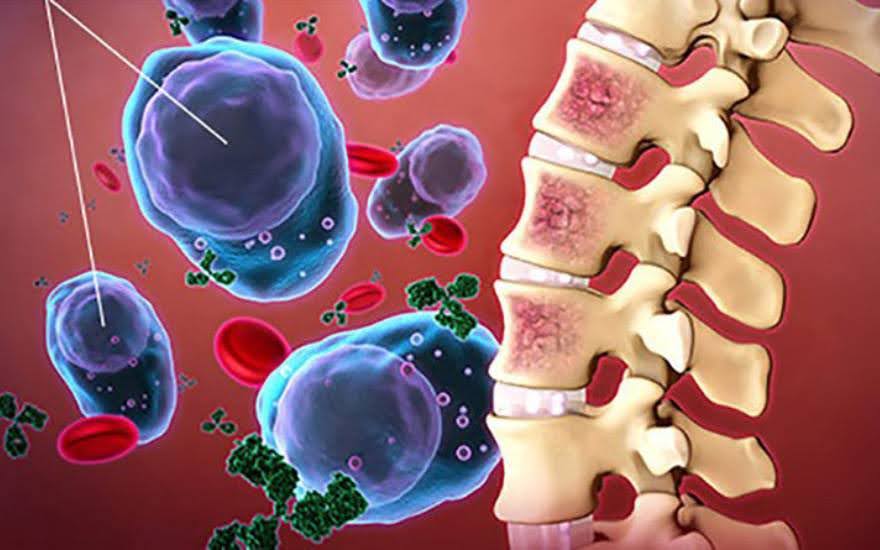
Plain radiography (X-ray) is typically the first imaging modality performed. Radiographic abnormalities such as periosteal reaction, cortical bone destruction, and soft tissue swelling usually appear only after two to four weeks from disease onset. In cases of spinal osteomyelitis, imaging may reveal reduced vertebral body height due to vertebral collapse, narrowing of the intervertebral disc space, or subchondral bone involvement.
When X-ray findings are inconclusive or early infection is suspected, computed tomography (CT) and magnetic resonance imaging (MRI) are preferred. These modalities allow early detection of bone involvement, accurate assessment of the extent of infection, and identification of associated complications such as abscess formation.
Treatment of osteomyelitis
Management of osteomyelitis involves two main approaches: medical therapy and surgical intervention, with the goals of eradicating infection and restoring bone integrity and function.
Medical management
- Antibiotic therapy plays a central role in the treatment of osteomyelitis. When culture results are positive, antibiotic selection is guided by antimicrobial susceptibility testing. The standard duration of antibiotic therapy is typically at least 4–6 weeks. In cases of chronic or recurrent osteomyelitis, treatment may be prolonged to 4–6 months, and in some instances up to 9 months. Staphylococcus aureus is the most common causative pathogen; therefore, anti-staphylococcal antibiotics such as vancomycin are often prioritized, particularly in patients with a history of prior medical or surgical intervention.
- Wound care is essential when there is associated skin infection or abscess formation. This includes meticulous wound cleansing, adequate drainage of abscesses, and, when indicated, continuous suction to remove infected exudate.
Surgical management
- Surgical debridement of infected tissueWhen antibiotic therapy alone is insufficient to eradicate infection, surgical intervention becomes necessary. This may involve thorough debridement of necrotic bone and infected soft tissue, or limb amputation in severe cases. Preoperative imaging studies, particularly magnetic resonance imaging (MRI), are used to accurately delineate the extent of infection. Postoperatively, close clinical monitoring is essential to promptly identify and manage any residual necrotic tissue or persistent infection.
- Bone reconstruction and replacementIn cases of extensive bone destruction, bone grafting or reconstructive procedures are required to replace the damaged bone and restore structural integrity. These techniques play a critical role in functional recovery and long-term stability of the affected limb.
- Removal and replacement of infected prosthetic jointsFor patients with prosthetic joint infection, removal of the infected implant is indicated. A two-stage revision arthroplasty, in which implant removal and reimplantation are performed in separate procedures, is generally preferred over a single-stage approach to reduce the risk of recurrent infection.
- Management of necrotic boneElimination of devitalized bone tissue and its replacement with healthy tissue is a key objective in osteomyelitis treatment. Whenever feasible, wounds should be closed early using local flaps, rotational flaps, or free tissue transfer to fill dead space, promote tissue regeneration, and restore normal bone anatomy and function.
The management of osteomyelitis requires close integration of medical and surgical approaches to achieve optimal outcomes and prevent long-term complications.
Osteomyelitis is a potentially serious condition that can cause significant tissue damage if not diagnosed and treated promptly. In addition, proactive prevention through good hygiene practices, effective control of underlying diseases, and maintenance of a healthy lifestyle plays an important role in protecting musculoskeletal health and improving overall quality of life.

Early detection and timely treatment are critical determinants in the effective control of osteomyelitis. Patients seeking evaluation and management of osteomyelitis at Hong Ngoc General Hospital are advised to contact the hotline 0889 621 046 to schedule an appointment and receive professional support from leading specialists.
Note: The information provided in this article by Hong Ngoc General Hospital is for reference purposes only and does not substitute for professional medical diagnosis or treatment. Patients should not self-medicate. For an accurate assessment of the condition, individuals are advised to visit a hospital or reputable healthcare facility for direct examination, diagnosis, and consultation regarding an appropriate treatment plan.
Follow the official fanpage of Hong Ngoc General Hospital for additional useful health information:https://www.facebook.com/Coxuongkhophongngoc