



A bone fracture is a condition in which the bone is broken or cracked due to strong impact from trauma or accidents. It causes pain, directly affects mobility, and requires timely medical intervention to restore function. So, how can fractures be accurately diagnosed and effectively treated?
Classification of bone fractures
There are several ways to classify bone fractures, such as by the condition of surrounding soft tissue (closed or open fractures) or by the characteristics of the fracture itself. Common classifications include:
- Closed fracture: A simple fracture without an open wound on the skin.
- Open fracture: Also called a compound fracture, in which the broken bone penetrates through the skin, creating an open wound. In some cases, the fractured bone may be exposed at the injury site.
- Complete fracture: The bone is broken into separate pieces. In severe cases, the bone may shatter into multiple fragments (comminuted fracture), displace from its original position, or even penetrate surrounding organs or the skin. This condition requires emergency care and prompt treatment.
- Incomplete fracture: The bone is only partially damaged without complete loss of continuity. Examples include hairline fractures or torus fractures (in which one side of the bone bends and bulges while the other side remains intact), commonly seen in children.
Symptoms of bone fracture
A bone fracture can often be recognized through the following symptoms:
- Visible deformity of the bone at the site of injury after trauma or accident
- Bruising around the injured area
- Swelling, redness, and severe pain that worsens with movement
- Loss of function in the affected area, such as inability to bend, stretch, or grip objects
- In cases of open fracture, the broken bone may pierce through the skin, causing bleeding and intense pain
Diagnostic methods for bone fractures
Bone fractures are diagnosed based on clinical symptoms combined with laboratory tests and imaging techniques:
- Clinical examination: Detects typical signs of fracture, helps classify the type of fracture, and guides the need for further imaging studies.
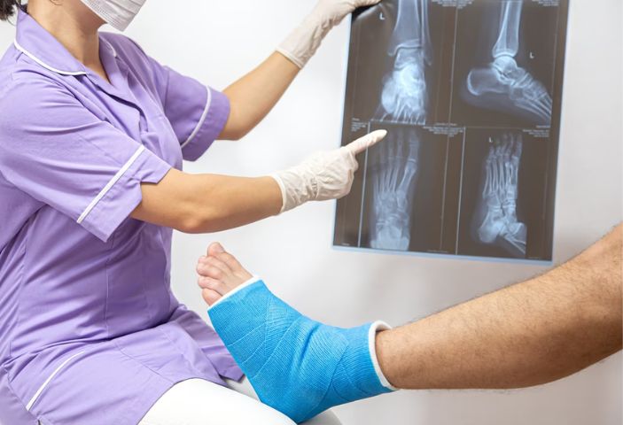
- Imaging techniques (X-ray, CT, MRI): Provide detailed two-dimensional or three-dimensional images of the bone, allowing physicians to accurately identify fracture lines, assess the extent of bone and joint damage, and evaluate surrounding tissues, muscles, tendons, and ligaments for associated injuries.
- Biochemical and hematological tests: Used to assess blood loss in fracture cases and detect potential wound infections, enabling physicians to predict outcomes and choose safe treatment approaches.
Complications after bone fracture
Some patients believe that a broken bone will heal on its own over time without medical intervention. In reality, fractures left untreated can lead to serious complications, including:
- Blood clots: Trauma may cause blood vessel blockage, reducing circulation and potentially leading to headaches, numbness, or muscle weakness.
- Compartment syndrome: When a bone fracture causes bleeding and swelling, tissue pressure inside the bone compartments increases, resulting in reduced blood supply. Without timely treatment, this can lead to rhabdomyolysis, hyperkalemia, infection, and, in the long term, muscle contracture, numbness, or paralysis of the affected area.
- Bone or joint infection: Bacteria may enter through open wounds and spread to the bone or bone marrow, causing persistent infections that are extremely difficult to treat and may last a lifetime.
- Bone malunion or deformity: Fractured bones may heal in the wrong position, resulting in deformity and reduced flexibility in movement.
- Nerve injury: Fracture fragments may compress or sever nearby nerves, causing sensory loss. In some cases, nerves cannot recover on their own and require timely surgical intervention.
Treatment methods for bone fractures
Depending on the severity of the injury, age, and medical history, physicians will determine the most appropriate treatment approach for each patient. The fundamental principles are to restore the fractured bone to its correct anatomical position, immobilize it, and promote the body’s natural bone healing process. Common treatment methods include:
- Casting Casts are typically made of plaster or fiberglass, providing firm protection that encases the fractured area and keeps the bone immobilized for several weeks, allowing natural healing. At Hong Ngoc General Hospital, lightweight fiberglass casts are applied only ⅛ the weight of traditional plaster casts. These specialized casts harden 20 times faster, helping to reduce pain quickly and significantly shorten treatment time. Made of waterproof fiberglass material, they allow patients to carry out daily activities, including bathing, without worrying about damage to the cast.
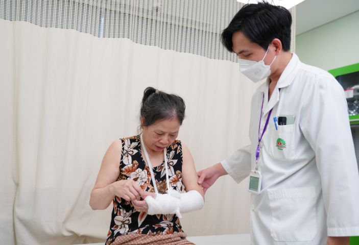
- Splinting This method is commonly used for closed fractures. The splint, often made of wood or other firm materials, is applied to one side of the fracture site to immobilize the bone, following the basic principles of fracture management.
- External fixation Metal pins or screws are inserted above and below the fracture site and then connected to an external metal frame. This system stabilizes the bone and prevents movement during the healing process.
- Skeletal traction A gentle, continuous pulling force is applied to the muscles and tendons around the fractured bone. This traction helps maintain proper bone alignment and supports the healing process.
- Internal fixation (biological osteosynthesis) A surgical method in which the fractured bone is repositioned and secured with screws, plates, or rods placed inside the body. This technique is indicated for complex fractures and ensures stability as well as precise bone reconstruction.
Reader may also be interested in:
- Anterior cruciate ligament (ACL) tear and optimal treatment approaches
- Meniscal tear of the knee: Causes and warning signs
Biological osteosynthesis surgery at Hong Ngoc General Hospital with Dr. Le Quang Huy (Orthopedic Trauma Surgery Specialist, European-trained) offers significant advantages for patients:
- Minimally invasive incision: Surgical incision of only 0.5–3 cm ensures small, aesthetic scars, reduced soft tissue damage, minimal blood loss, preservation of blood supply at the fracture site, and maximum reduction of infection risk.
- Safe and biocompatible implants: Screws and plates made from biocompatible materials provide strong stability during movement, stimulate the formation of bone cells, cartilage cells, and collagen fibers, enabling natural bone healing and allowing patients to regain mobility as early as one day after surgery.
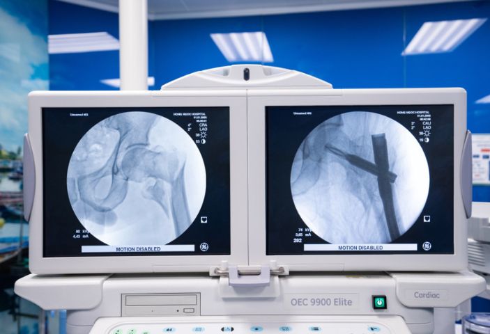
- Advanced medical equipment: State-of-the-art orthopedic surgical tables and C-arm imaging systems help accurately identify fracture sites, restore correct bone alignment, and minimize postoperative complications.
Note: After the initial treatment phase, patients may be advised to undergo physical therapy targeting the fractured area to restore mobility, prevent muscle atrophy, and avoid joint stiffness.
For more in-depth consultation on fracture treatment at Hong Ngoc General Hospital, please contact our hotline at 0912002131 or 0949646556 for timely support.
Note: The information provided in this article by Hong Ngoc General Hospital is for reference only and is not a substitute for professional medical diagnosis or treatment. Patients should not attempt self-treatment. For an accurate assessment of your condition, please visit the hospital for direct examination, diagnosis, and a personalized treatment plan from a physician.
Follow the official fanpage of Hong Ngoc General Hospital for more useful health information: https://www.facebook.com/BenhvienHongNgoc