Varicose veins may initially present with localized pain or discomfort along the affected veins. While the condition is not typically associated with severe symptoms in its early stages, delayed management can lead to venous thrombosis, a potentially serious complication that may pose significant health risks and, in severe cases, become life-threatening.
What are varicose veins?
Veins are responsible for transporting deoxygenated blood from peripheral tissues back to the heart to initiate the next cycle of circulation. Venous insufficiency and varicosities can occur in any anatomical location; however, they most commonly affect the lower extremities due to their distance from the heart and the sustained hydrostatic pressure exerted by body weight.
Varicose veins are characterized by venous blood stasis, leading to increased intraluminal pressure and progressive venous dilation. As venous dilation worsens over time, arterial perfusion to the lower limbs may be adversely affected.
In the lower extremities, varicosities may involve either the superficial or deep venous systems, resulting in dilated, tortuous veins associated with pain, heaviness, and fatigue.
Causes of varicose veins
Varicose veins are primarily caused by inflammation of the venous wall and venous reflux, in which blood flows backward toward the lower limbs. This process impairs the return of blood from the legs to the heart, leading to circulatory stasis. Over time, the veins progressively dilate and enlarge, potentially resulting in complications such as chronic venous insufficiency and venous thrombosis.
Risk factors for varicose veins include:
- A family history of varicose veins
- Higher prevalence in females compared to males
- Overweight and obesity
- Advanced age, with a higher risk observed in older adults than in younger individuals
- Pregnancy, as the enlarging uterus exerts pressure on abdominal vessels, increasing venous pressure in the lower extremities and contributing to venous dilation
- Occupations requiring prolonged standing, such as teachers, sales staff, and similar professions
Individuals with the aforementioned risk factors have a higher likelihood of developing varicose veins compared to the general population.
Recognizing the symptoms of varicose veins
In the early stages, varicose veins are often asymptomatic or present with subtle, nonspecific symptoms that are difficult to recognize. Patients may experience mild heaviness, discomfort, a sensation of warmth, and itching in the legs, particularly after prolonged walking or increased physical activity involving the lower limbs.
These symptoms typically become more pronounced toward the end of the day or after prolonged standing. Patients may also report paresthesia, described as tingling or a “pins and needles” sensation in the calves, and may experience frequent muscle cramps.
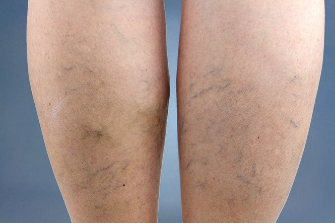
In more advanced stages, clinical manifestations become more apparent. Patients may observe small, dilated superficial vessels visible on the skin surface. In cases where venous dilation is not severe, these signs may subside after rest.
Because the symptoms of varicose veins are often subtle and may resolve with rest, they are easily overlooked. However, for accurate evaluation, individuals should seek cardiovascular assessment if the following symptoms are present:
- Persistent leg fatigue, with a sensation of tightness or heaviness in the calves
- Frequent muscle cramps and paresthesia (tingling or “pins and needles”), particularly at night
- Inflammation of superficial veins, visible as bluish veins in the thigh, ankle, or knee regions
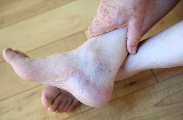
- Swelling and pruritus of the legs, especially around the ankles
- Skin changes in the lower limbs, including discoloration and soft tissue infection near the ankle area
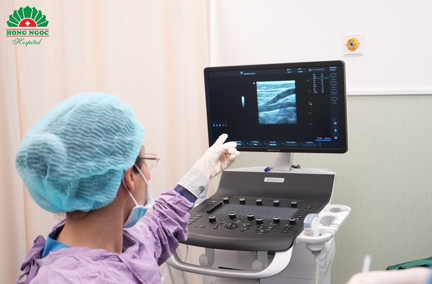
During clinical evaluation, Doppler ultrasound of the vascular system enables physicians to accurately diagnose the presence of varicose veins.
Are varicose veins dangerous?
Varicose veins do not typically cause severe symptoms in the early stages. However, without appropriate treatment, the condition may lead to serious complications.
In addition to symptoms such as pain, discomfort, pruritus, and muscle cramps, the presence of thrombus formation near varicose veins can significantly worsen clinical manifestations. In particular, deep vein thrombosis (DVT) may be life-threatening due to significant obstruction of blood flow.
Superficial venous thrombosis is generally less dangerous; however, if secondary infection occurs in the surrounding tissues, prompt medical intervention is required. Notably, a thrombus may dislodge and migrate to the lungs, resulting in pulmonary embolism, a potentially fatal condition if not treated in a timely manner.
Reader may also be interested in:
- Aortic aneurysm and treatment options
- Deep vein thrombosis of the lower extremities: early treatment to prevent serious complications
In pregnant women, early management of varicose veins is essential. Those with coagulation disorders or reduced mobility are at increased risk of venous thrombosis. Therefore, careful monitoring is recommended if symptoms such as thigh or leg swelling, pain, or low-grade fever occur.
How are varicose veins diagnosed?
To diagnose varicose veins, physicians first obtain a detailed medical history, including the patient’s overall health status, presenting symptoms, and relevant risk factors. This is followed by a clinical examination, during which the physician assesses the color, morphology, and prominence of superficial veins.
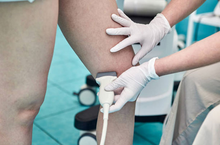
To diagnose varicose veins, Doppler ultrasound is commonly indicated. This imaging modality utilizes high-frequency sound waves to assess blood flow velocity within the veins and evaluate venous structure.
In addition to accurately identifying venous dilation, Doppler ultrasound also helps determine underlying causes, including conditions not primarily originating from the venous system. This enables clinicians to establish an appropriate and effective treatment strategy.
Treatment of varicose veins
Varicose veins can be managed using a range of treatment modalities, depending on the severity and clinical progression of the disease. Commonly employed approaches include:
Medical management
In mild cases, physicians may prescribe pharmacological therapy. Medications help reduce venous reflux and improve venous return. Venoactive agents (phlebotonics) may be used to enhance venous wall tone; however, their effectiveness is primarily limited to the early stages of the disease.
Surgical treatment
Surgical intervention involves the removal of dilated superficial veins using specialized instruments. This approach offers high efficacy and a low recurrence rate. However, it is now less commonly performed due to the need for anesthesia, prolonged hospitalization, delayed postoperative recovery, and potential complications such as thigh hematoma, venous thrombosis, and lower limb paresthesia.
Cryotherapy with liquid nitrogen (−90°C)This technique occludes the venous lumen using an intravascular catheter combined with extreme cold. Its main limitation is a relatively high recurrence rate, which can reach up to 30%.
Sclerotherapy
This method involves injecting a sclerosing agent into the superficial venous system of the lower extremities. The agent induces endothelial damage and affects the adjacent medial layer, leading to thrombus formation and subsequent occlusion of the incompetent vein. As a result, blood no longer pools within the dilated vein.
Endovenous interventions
These minimally invasive techniques use laser or radiofrequency energy to ablate the affected vein. Endovenous laser therapy (EVLT) employs fiber-optic catheters delivering laser energy, commonly at a wavelength of 1470 nm, while radiofrequency ablation (RFA) utilizes catheters that emit high-frequency energy to achieve venous closure.
Both techniques generate thermal energy that destroys collagen within the venous wall, leading to endothelial damage, inflammation, fibrosis, and complete occlusion of the affected vein.
Endovenous ablation using radiofrequency or laser is an advanced technique that requires a high level of clinical expertise and modern medical equipment.
At Hong Ngoc General Hospital, radiofrequency ablation (RFA) for the treatment of lower extremity varicose veins has been successfully implemented, with favorable outcomes in a large number of patients, significantly alleviating symptoms and improving quality of life.
Register for consultation and expert evaluation here:
Note: The information provided in this article by Hong Ngoc General Hospital is for reference purposes only and does not replace professional medical diagnosis or treatment. Patients should not self-medicate. For an accurate assessment of their condition, individuals are advised to visit a medical facility for direct examination, diagnosis, and appropriate treatment planning by qualified physicians.
Follow the official fanpage of Hong Ngoc General Hospital for more health-related information and updates.