Cholecystitis is one of the most common digestive complications, particularly in middle-aged and older adults. If left untreated, it can lead to serious consequences, with surgical removal of the gallbladder often being the necessary intervention.
What is cholecystitis?
The gallbladder is a small, pear-shaped organ located on the right side of the abdomen, beneath the liver. It stores bile, a digestive fluid produced by the liver, which is released into the small intestine to help break down fats during digestion.
Cholecystitis is an infection or inflammation of the gallbladder. If not treated promptly, the condition may progress to serious complications, including gallbladder perforation.
Symptoms of cholecystitis
Cholecystitis may present in two forms:
- Acute cholecystitis (sudden onset):
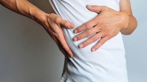
- Severe pain in the upper right abdomen
- Pain radiating to the right shoulder or back
- In severe cases, gallbladder perforation may occur, causing bile leakage into the abdominal cavity. This leads to intense pain and can be life-threatening, requiring immediate emergency care
- Additional symptoms may include nausea, vomiting, and fever
- Chronic cholecystitis (recurrent inflammation over months or years):
- Nausea
- Vomiting
- Fever
- Pain in the upper right abdomen
- Mid-abdominal tenderness on palpation
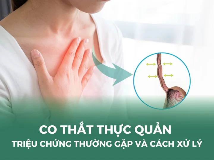
Symptoms of cholecystitis are usually most frequent and severe after meals, as the gallbladder contracts to release bile into the small intestine to aid digestion.
Causes of cholecystitis
Cholecystitis may develop due to several underlying factors, including:
- Gallstones: The most common cause of cholecystitis. Gallstones are hardened deposits that form inside the gallbladder. They can block the cystic duct, preventing bile from flowing out, which leads to bile buildup and inflammation.
- Infection: Certain infections, including viral illnesses or conditions such as AIDS, can trigger gallbladder inflammation.
- Tumors: A tumor may compress or obstruct the bile ducts, preventing bile from draining and resulting in gallbladder inflammation.
- Other causes: Vascular problems that reduce blood flow to the gallbladder may also contribute to cholecystitis. In addition, scarring within the bile ducts may obstruct bile flow and cause inflammation.
Who is at risk of developing cholecystitis?
Certain groups of people have a higher risk of developing cholecystitis compared with the general population, including:
- Individuals in middle age or older
- People with obesity or those who consume a diet high in fat
- Individuals who frequently fast, leaving the stomach and intestines empty for extended periods
- Patients experiencing side effects from certain specialized medications
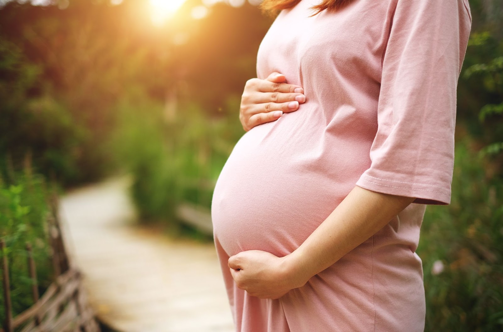
- Pregnant women, who face a higher risk compared with non-pregnant women
Is cholecystitis dangerous?
If left untreated, cholecystitis can lead to several serious complications, including:
- Gallbladder infection: Accumulated bile can become infected, worsening the inflammation.
- Gallbladder necrosis: Prolonged untreated cholecystitis may cause tissue death within the gallbladder. This is one of the most common complications, particularly in older adults and patients with diabetes.
- Gallbladder perforation: Severe swelling, infection, or necrosis may cause the gallbladder wall to rupture.
- Gallbladder cancer: Chronic or recurrent inflammation of the gallbladder may increase the risk of gallbladder carcinoma.
Reader may also be interested in:
- Are gallstones dangerous? Treatment options for gallstones
- Common bile duct stones: Causes, complications, and treatment
Diagnosis of cholecystitis
Several diagnostic methods are commonly used to confirm cholecystitis and assess its severity:
- Blood tests: Blood tests help determine whether an infection is present in the gallbladder. An elevated white blood cell count is often a sign of infection. Biochemical tests are also performed to evaluate kidney and liver function.
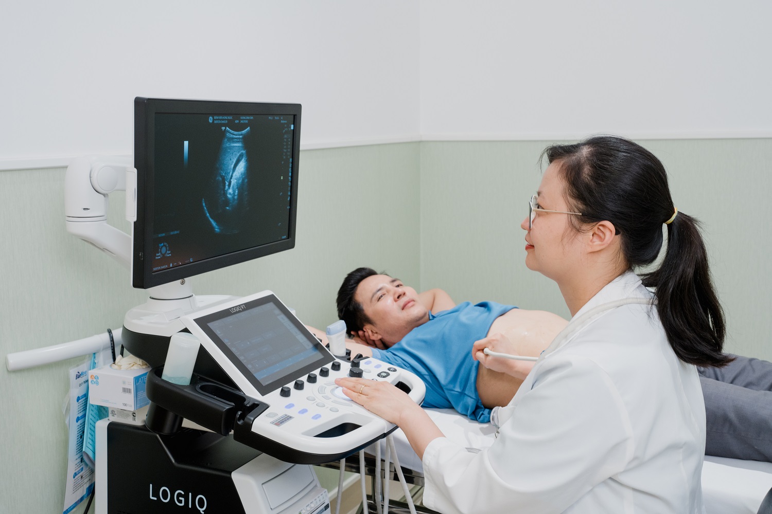
- Abdominal ultrasound: Ultrasound uses sound waves to create images of the gallbladder and bile ducts. It is the primary imaging tool for detecting signs of gallbladder inflammation and can clearly reveal gallstones.
- Cholecystography (gallbladder X-ray): By administering a contrast agent orally or intravenously, X-ray imaging of the abdomen can be performed to evaluate disorders of the liver, gallbladder, and bile ducts. In acute cholecystitis, this method may detect bile duct obstruction.
- Computed tomography (CT scan): A CT scan uses X-rays to generate detailed images of the abdomen, including the liver, gallbladder, bile ducts, and intestines. It can help identify gallbladder inflammation, bile flow obstruction, or occasionally gallstones.
- Magnetic resonance cholangiopancreatography (MRCP): MRCP provides detailed images of the liver, gallbladder, bile ducts, pancreas, and pancreatic ducts. It is particularly useful in detecting gallstones, cholecystitis, cholangitis, or bile flow obstruction.
Treatment of cholecystitis
Medical (non-surgical) management
- Pain relief: Analgesics are prescribed to control abdominal pain.
- Antibiotics: Used to treat or prevent gallbladder infection.
- Dietary modification: A low-fat diet with smaller, more frequent meals is recommended to reduce strain on the gallbladder.
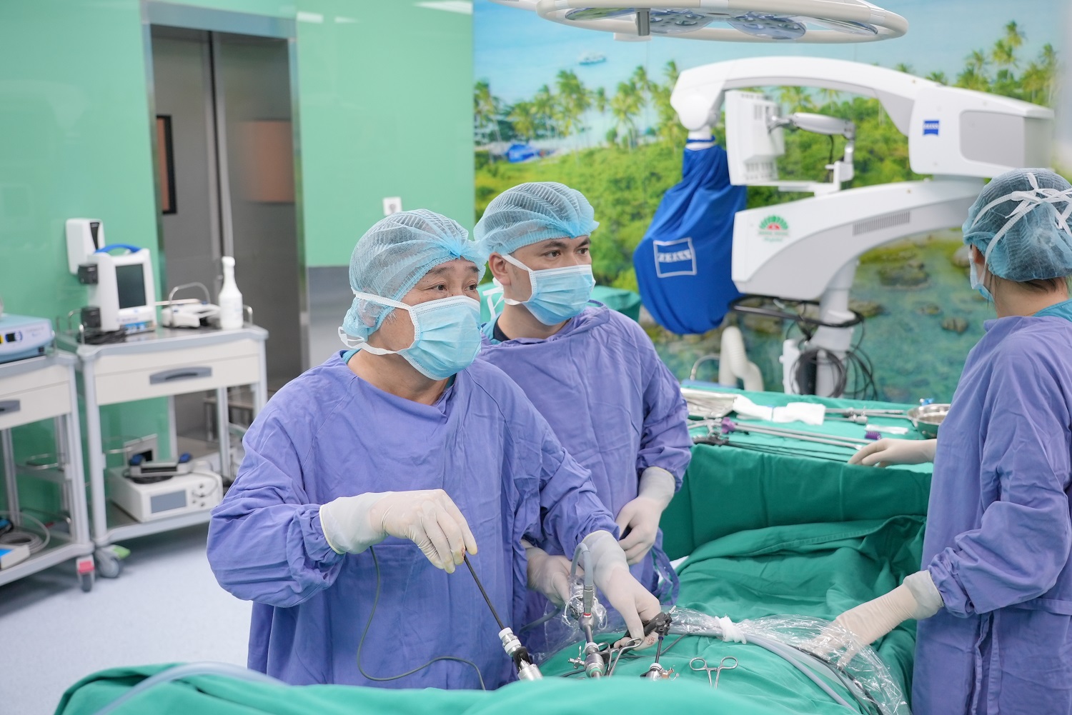
Surgical management
- Laparoscopic cholecystectomy: The most common procedure, performed under general anesthesia, in which the gallbladder is removed through minimally invasive keyhole surgery.
- Open cholecystectomy: In certain cases, an abdominal incision is made to remove the gallbladder under general anesthesia.
Alternative procedures (for patients not suitable for surgery, particularly in cases of gallstone-related obstruction):
- Percutaneous cholecystolithotomy: A minimally invasive approach to break up and remove gallstones through the skin.
- Endoscopic retrograde cholangiopancreatography (ERCP): A combined endoscopic and radiographic technique to identify and remove stones obstructing the bile ducts.
- Percutaneous transhepatic cholangiography (PTC): An interventional imaging method used to visualize and relieve bile duct obstruction.
Note: The information provided in this article by Hong Ngoc General Hospital is for reference only and is not a substitute for professional medical diagnosis or treatment. Patients should not self-medicate. For an accurate assessment of the condition, it is essential to visit a hospital for direct examination, diagnosis, and consultation with a physician to receive an appropriate treatment plan and the most effective prescription.
Gastroenterology Center – Hong Ngoc General Hospital
- Hong Ngoc Phuc Truong Minh General Hospital – No. 8 Chau Van Liem, Tu Liem, Hanoi
- Hong Ngoc General Hospital – 55 Yen Ninh, Ba Dinh, Hanoi
- Hong Ngoc Savico Long Bien Clinic – 3rd Floor, Block B, Tasco Megamall, 07–09 Viet Hung, Hanoi
Hotline: 0911 908 856
>>> Stay updated with useful health information and special offers at: https://www.facebook.com/trungtamtieuhoaBVHongNgoc