Pulmonary consolidation syndrome in children poses significant risks if not detected and treated promptly. A proper understanding of the condition, along with early recognition of warning signs, can help parents prevent long-term lung damage and minimize complications that may affect a child’s overall growth and development.
What is pulmonary consolidation in children?
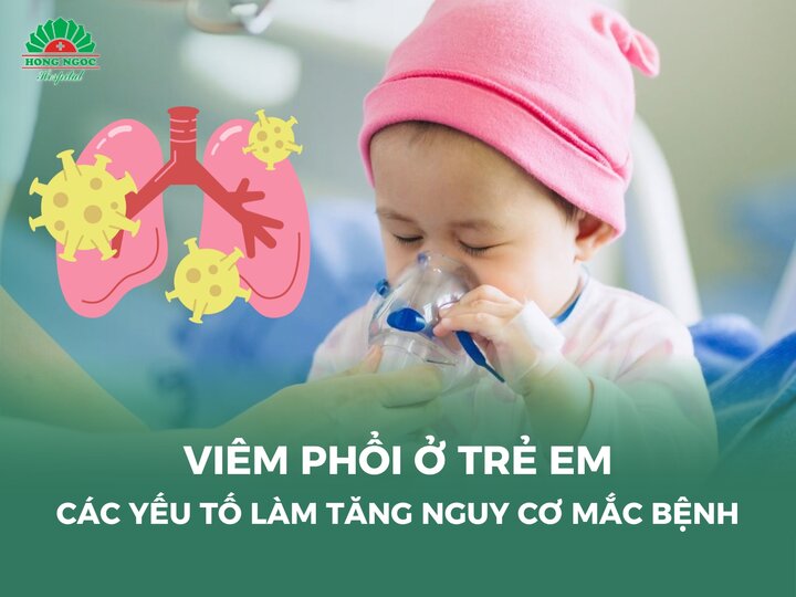
Pulmonary consolidation in children refers to a condition in which the normal air-filled lung tissue is replaced by fluid (such as pus, plasma, or blood), inflammatory cells, or abnormal secretions. This process leads to loss of elasticity and impaired ventilation of the lung. As a result, the consolidated lung areas are no longer able to perform effective gas exchange, causing hypoxemia and characteristic clinical manifestations.
This syndrome is most often a consequence of underlying conditions such as pneumonia, pulmonary tuberculosis, pulmonary infarction, or pulmonary edema, all of which directly affect the lung parenchyma or disrupt the process of ventilation and gas exchange.
Causes of pulmonary consolidation syndrome in children
Pneumonia
Pneumonia is the most common cause, in which inflammatory exudates, pus, and necrotic cells fill the small airways and alveoli.
Acute lobar pneumonia: Often caused by Streptococcus pneumoniae or atypical bacteria such as Mycoplasma pneumoniae.
Bacterial pneumonia from aerobic or anaerobic pathogens: Infections due to Haemophilus influenzae, Staphylococcus aureus, or Gram-negative bacteria such as Klebsiella pneumoniae may result in severe pneumonia with extensive pulmonary consolidation.
Tuberculous pneumonia: Mycobacterium tuberculosis induces granulomatous lesions that may progress to pulmonary tuberculosis, frequently causing consolidation in the upper or middle lobes.
Lung abscess
Lung abscesses typically develop when anaerobic bacteria (e.g., Bacteroides spp.) or aerobic bacteria (Staphylococcus aureus) infect the lung parenchyma, leading to the formation of pus-filled cavities. The affected lung area becomes consolidated due to the accumulation of pus and necrotic tissue within the alveoli. Risk factors include aspiration, immunosuppression, or complications arising from untreated pneumonia.
Atelectasis due to bronchial obstruction
Enlarged lymph nodes (as in primary tuberculosis or malignant disease) or intrapulmonary tumors may compress a main bronchus, causing partial lung collapse. Airway obstruction due to a foreign body or blood clot can also lead to this condition. When a lung lobe is deprived of air, the alveoli collapse and lose their air content, resulting in consolidation.
Pulmonary infarction (due to pulmonary artery obstruction)
Blockage of the pulmonary artery may be caused by:
Thromboembolism from deep vein thrombosis
Air embolism (post-surgical or secondary to negative intrathoracic pressure)
Fat embolism (after long-bone fracture) Associated conditions include cardiac diseases (mitral stenosis, heart failure) or hypercoagulable states (thrombophilia, postpartum women). When blood flow to a lung segment is obstructed, tissue ischemia ensues, leading to parenchymal injury and localized atelectasis.
Less common causes
Pulmonary edema: Often occurs in left-sided heart failure or acute respiratory distress syndrome (ARDS), resulting in alveolar fluid accumulation and consolidation.
Lung cancer: Primary tumors or metastatic lymph nodes compressing the bronchus can cause localized atelectasis and consolidation.
Trauma or lung injury: Aspiration of gastric contents, chemical inhalation, or blunt chest trauma may induce chemical pneumonitis and subsequent consolidation.
Pneumonia is the most common underlying cause of pulmonary consolidation syndrome in children.
Clinical manifestations of pulmonary consolidation in children
The common symptoms of pulmonary consolidation in children, regardless of the underlying cause, often include:
High fever, chills, excessive sweating (especially at night), fatigue, and physical exhaustion.
Pallor or cyanosis of the skin and lips.
Respiratory distress, wheezing, and increasing shortness of breath as the extent of consolidation worsens.
Rapid or shallow breathing, difficulty speaking, or shortness of breath during exertion.
Chest pain or a feeling of heaviness in the chest.
Cough, which may be mild or severe, often productive of sputum, and occasionally accompanied by hemoptysis.
Children with pulmonary consolidation syndrome often present with high fever and chills.
Serious complications of pulmonary consolidation in children
Acute respiratory failure: When lung parenchyma becomes consolidated, the ability of alveoli to perform gas exchange is markedly reduced or completely lost. This results in hypoxemia and hypercapnia, progressing to acute respiratory failure.
Sepsis and septic shock: Severe pneumonia or lung abscess may allow bacteria to enter the bloodstream, causing sepsis. This condition can lead to multi-organ dysfunction and carries a high risk of mortality if not treated promptly.
Pleural effusion or empyema: Infection from the lung parenchyma can spread to the pleural space, resulting in the accumulation of inflammatory fluid or pus. The effusion compresses the lung, further impairing its ability to expand and worsening consolidation.
Atelectasis: Prolonged consolidation, combined with bronchial obstruction due to mucus plugs or external compression, may cause partial or complete collapse of a lung lobe.
Bronchiectasis: Chronic infection and inflammation within the lung parenchyma damage and destroy bronchial wall structures, leading to irreversible bronchiectasis.
Chronic lung abscess formation: Untreated or recurrent lung abscesses may evolve into chronic infectious foci, significantly compromising lung function.
Note: The information provided in this article by Hong Ngoc General Hospital is for reference only and does not replace professional medical diagnosis or treatment. To determine the exact condition, parents should take their child to the hospital for direct examination and receive an appropriate treatment plan from a physician.
With the motto “Safe pediatric care – preventing cross-infection, avoiding antibiotic overuse, and minimizing recurrence,” Hong Ngoc Pediatrics Department takes pride in being a trusted destination for parents seeking quality medical examination and treatment for their children.
HONG NGOC PEDIATRICS DEPARTMENT – HONG NGOC GENERAL HOSPITAL
2 hospital locations:
55 Yen Ninh, Ba Dinh, Hanoi
No. 8 Chau Van Liem, Nam Tu Liem, Hanoi
5 polyclinic locations:
No. 7 & 9 Nguyen Van Linh Street, Viet Hung, Hanoi
1st Floor, HPC Landmark 105, Ha Dong, Hanoi
1st Floor, Autumn Building – Gold Season Urban Area, No. 47 Nguyen Tuan, Thanh Xuan, Hanoi
1st & 2nd Floors, NoVo Building, Kosmo Apartment Complex, 161 Xuan La, Xuan Dinh, Hanoi
10th Floor, Keangnam Hanoi Landmark Tower, Yen Hoa, Hanoi
Free consultation and fast appointment booking hotline: 0947 616 006
Pulmonary consolidation syndrome in children poses significant risks if not detected and treated promptly. A proper understanding of the condition, along with early recognition of warning signs, can help parents prevent long-term lung damage and minimize complications that may affect a child’s overall growth and development.
What is pulmonary consolidation in children?
Pulmonary consolidation in children refers to a condition in which the normal air-filled lung tissue is replaced by fluid (such as pus, plasma, or blood), inflammatory cells, or abnormal secretions. This process leads to loss of elasticity and impaired ventilation of the lung. As a result, the consolidated lung areas are no longer able to perform effective gas exchange, causing hypoxemia and characteristic clinical manifestations.
This syndrome is most often a consequence of underlying conditions such as pneumonia, pulmonary tuberculosis, pulmonary infarction, or pulmonary edema, all of which directly affect the lung parenchyma or disrupt the process of ventilation and gas exchange.
Causes of pulmonary consolidation syndrome in children
Pneumonia
Pneumonia is the most common cause, in which inflammatory exudates, pus, and necrotic cells fill the small airways and alveoli.
Acute lobar pneumonia: Often caused by Streptococcus pneumoniae or atypical bacteria such as Mycoplasma pneumoniae.
Bacterial pneumonia from aerobic or anaerobic pathogens: Infections due to Haemophilus influenzae, Staphylococcus aureus, or Gram-negative bacteria such as Klebsiella pneumoniae may result in severe pneumonia with extensive pulmonary consolidation.
Tuberculous pneumonia: Mycobacterium tuberculosis induces granulomatous lesions that may progress to pulmonary tuberculosis, frequently causing consolidation in the upper or middle lobes.
Lung abscess
Lung abscesses typically develop when anaerobic bacteria (e.g., Bacteroides spp.) or aerobic bacteria (Staphylococcus aureus) infect the lung parenchyma, leading to the formation of pus-filled cavities. The affected lung area becomes consolidated due to the accumulation of pus and necrotic tissue within the alveoli. Risk factors include aspiration, immunosuppression, or complications arising from untreated pneumonia.
Atelectasis due to bronchial obstruction
Enlarged lymph nodes (as in primary tuberculosis or malignant disease) or intrapulmonary tumors may compress a main bronchus, causing partial lung collapse. Airway obstruction due to a foreign body or blood clot can also lead to this condition. When a lung lobe is deprived of air, the alveoli collapse and lose their air content, resulting in consolidation.
Pulmonary infarction (due to pulmonary artery obstruction)
Blockage of the pulmonary artery may be caused by:
Thromboembolism from deep vein thrombosis
Air embolism (post-surgical or secondary to negative intrathoracic pressure)
Fat embolism (after long-bone fracture) Associated conditions include cardiac diseases (mitral stenosis, heart failure) or hypercoagulable states (thrombophilia, postpartum women). When blood flow to a lung segment is obstructed, tissue ischemia ensues, leading to parenchymal injury and localized atelectasis.
Less common causes
Pulmonary edema: Often occurs in left-sided heart failure or acute respiratory distress syndrome (ARDS), resulting in alveolar fluid accumulation and consolidation.
Lung cancer: Primary tumors or metastatic lymph nodes compressing the bronchus can cause localized atelectasis and consolidation.
Trauma or lung injury: Aspiration of gastric contents, chemical inhalation, or blunt chest trauma may induce chemical pneumonitis and subsequent consolidation.
Pneumonia is the most common underlying cause of pulmonary consolidation syndrome in children.
Clinical manifestations of pulmonary consolidation in children
The common symptoms of pulmonary consolidation in children, regardless of the underlying cause, often include:
High fever, chills, excessive sweating (especially at night), fatigue, and physical exhaustion.
Pallor or cyanosis of the skin and lips.
Respiratory distress, wheezing, and increasing shortness of breath as the extent of consolidation worsens.
Rapid or shallow breathing, difficulty speaking, or shortness of breath during exertion.
Chest pain or a feeling of heaviness in the chest.
Cough, which may be mild or severe, often productive of sputum, and occasionally accompanied by hemoptysis.
Children with pulmonary consolidation syndrome often present with high fever and chills.
Serious complications of pulmonary consolidation in children
Acute respiratory failure: When lung parenchyma becomes consolidated, the ability of alveoli to perform gas exchange is markedly reduced or completely lost. This results in hypoxemia and hypercapnia, progressing to acute respiratory failure.
Sepsis and septic shock: Severe pneumonia or lung abscess may allow bacteria to enter the bloodstream, causing sepsis. This condition can lead to multi-organ dysfunction and carries a high risk of mortality if not treated promptly.
Pleural effusion or empyema: Infection from the lung parenchyma can spread to the pleural space, resulting in the accumulation of inflammatory fluid or pus. The effusion compresses the lung, further impairing its ability to expand and worsening consolidation.
Atelectasis: Prolonged consolidation, combined with bronchial obstruction due to mucus plugs or external compression, may cause partial or complete collapse of a lung lobe.
Bronchiectasis: Chronic infection and inflammation within the lung parenchyma damage and destroy bronchial wall structures, leading to irreversible bronchiectasis.
Chronic lung abscess formation: Untreated or recurrent lung abscesses may evolve into chronic infectious foci, significantly compromising lung function.
Note: The information provided in this article by Hong Ngoc General Hospital is for reference only and does not replace professional medical diagnosis or treatment. To determine the exact condition, parents should take their child to the hospital for direct examination and receive an appropriate treatment plan from a physician.
With the motto “Safe pediatric care – preventing cross-infection, avoiding antibiotic overuse, and minimizing recurrence,” Hong Ngoc Pediatrics Department takes pride in being a trusted destination for parents seeking quality medical examination and treatment for their children.
HONG NGOC PEDIATRICS DEPARTMENT – HONG NGOC GENERAL HOSPITAL
2 hospital locations:
55 Yen Ninh, Ba Dinh, Hanoi
No. 8 Chau Van Liem, Nam Tu Liem, Hanoi
5 polyclinic locations:
No. 7 & 9 Nguyen Van Linh Street, Viet Hung, Hanoi
1st Floor, HPC Landmark 105, Ha Dong, Hanoi
1st Floor, Autumn Building – Gold Season Urban Area, No. 47 Nguyen Tuan, Thanh Xuan, Hanoi
1st & 2nd Floors, NoVo Building, Kosmo Apartment Complex, 161 Xuan La, Xuan Dinh, Hanoi
10th Floor, Keangnam Hanoi Landmark Tower, Yen Hoa, Hanoi
Free consultation and fast appointment booking hotline: 0947 616 006
Để lại câu hỏi của bạn để nhận được giải đáp từ các bác sĩ của Hồng Ngọc
Lorem ipsum dolor
Lorem ipsum dolor sit amet, consectetur adipiscing elit. Mauris odio lectus, pretium faucibus nisi eu, accumsan consectetur orci. In blandit vehicula nisl, vel lacinia ligula finibus a. Donec fermentum rhoncus
Lorem ipsum dolor sit amet, consectetur adipiscing elit. Mauris odio lectus, pretium faucibus nisi eu, accumsan consectetur orci. In blandit vehicula nisl, vel lacinia ligula finibus a. Donec fermentum rhoncus
Lorem ipsum dolor sit amet, consectetur adipiscing elit. Mauris odio lectus, pretium faucibus nisi eu, accumsan consectetur orci. In blandit vehicula nisl, vel lacinia ligula finibus a. Donec fermentum rhoncus
Lorem ipsum dolor sit amet, consectetur adipiscing elit. Mauris odio lectus, pretium faucibus nisi eu, accumsan consectetur orci. In blandit vehicula nisl, vel lacinia ligula finibus a. Donec fermentum rhoncus
Lorem ipsum dolor sit amet, consectetur adipiscing elit. Mauris odio lectus, pretium faucibus nisi eu, accumsan consectetur orci. In blandit vehicula nisl, vel lacinia ligula finibus a. Donec fermentum rhoncus