



Peripartum cardiomyopathy is a form of idiopathic dilated cardiomyopathy. This condition is initiated by a cascade of inflammatory processes that result in myocardial injury and subsequent heart failure. It typically occurs in the late stages of pregnancy or, more commonly, within the early postpartum period. Although it can be challenging to diagnose, the condition is potentially treatable if identified and managed promptly.
What is peripartum cardiomyopathy?
Peripartum cardiomyopathy (PPCM) is a rare form of heart failure characterized by dilation of the cardiac chambers and impaired myocardial contractility. This results in a reduced ejection fraction and decreased cardiac output with each heartbeat.
This condition affects women during the late stages of pregnancy and is most commonly diagnosed within the first five months postpartum. Peripartum cardiomyopathy is more frequently observed in women over the age of 30.
.jpg)
Epidemiology of peripartum cardiomyopathy
Peripartum cardiomyopathy is a relatively rare condition. It is estimated that at least 1 in 50,000 pregnant women may develop this disorder. Epidemiological studies have shown that women of African and Asian descent have a higher risk of developing peripartum cardiomyopathy.
In the United States, the incidence is approximately 1 in 1,000 pregnancies, with higher prevalence reported in the southern regions compared to other areas.
Notably, the condition tends to affect Black women more severely than White women and is often diagnosed at a younger age, typically over 27 years. In many cases, the disease is detected at a late stage, when significant myocardial damage has already occurred.
How is peripartum cardiomyopathy diagnosed?
Peripartum cardiomyopathy can be challenging to diagnose, as its clinical manifestations often overlap with normal physiological changes during pregnancy. Therefore, any occurrence of dyspnea or edema that develops suddenly during pregnancy or persists after delivery should be promptly evaluated by a healthcare professional.
Diagnostic criteria for peripartum cardiomyopathy
Peripartum cardiomyopathy is diagnosed based on the following key clinical criteria:
- Heart failure developing in the last month of pregnancy or within several months postpartum
- Reduced left ventricular systolic function, with a left ventricular ejection fraction (LVEF) < 45 percent, typically assessed by echocardiography (normal LVEF ranges from 50 to 70 percent)
- Absence of any other identifiable cause of heart failure
Diagnostic evaluation
Initial assessment through clinical examination and laboratory testing plays a crucial role in identifying abnormalities in renal function, electrolyte balance, hepatic function, and thyroid status. Patients with peripartum cardiomyopathy often exhibit elevated levels of brain natriuretic peptide (BNP) and N-terminal pro–BNP.
- Chest radiography may reveal cardiomegaly and/or pulmonary edema.
- Electrocardiography (ECG/EKG) is used to detect cardiac arrhythmias and other electrical abnormalities.
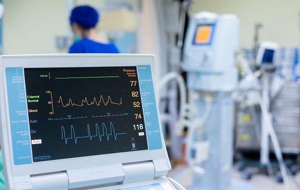
- Echocardiography is performed to assess cardiac anatomy and function. It also helps identify other potential causes of heart failure, such as valvular heart disease or structural abnormalities. In addition, echocardiography allows evaluation of left ventricular ejection fraction (LVEF), which is essential for the diagnosis of peripartum cardiomyopathy.
- Cardiac magnetic resonance imaging (MRI) is used to exclude other causes of heart failure and to evaluate chamber volumes and ventricular function.
- Cardiac catheterization may be indicated in patients with suspected ischemic peripartum cardiomyopathy.
- Endomyocardial biopsy is not routinely recommended and is primarily reserved for evaluating infectious causes of heart failure.
For cardiovascular consultation, please register here:
Severity and risks in pregnant and postpartum women
The severity of peripartum cardiomyopathy varies between pregnant and postpartum women. In some severe cases, heart transplantation may be required, with approximately 9 percent mortality related to postoperative complications.
Pregnant women
Pregnant women with peripartum cardiomyopathy are often indicated for cesarean delivery, and there is an increased risk of preterm birth and low birth weight. The recurrence rate in subsequent pregnancies is approximately 30 percent.
Close monitoring by healthcare professionals is essential. If the patient’s condition remains stable, vaginal delivery may still be considered. In some well-managed cases, cardiac function can recover to normal after delivery.
Postpartum women
If the left ventricular ejection fraction (LVEF) is less than 30 percent at the time of diagnosis, patients are at high risk of severe complications.
In cases where no improvement is observed within five years, patients may develop chronic heart failure and congestive complications.
The mortality rate may reach up to 50 percent in patients with delayed diagnosis of peripartum cardiomyopathy.
Causes of peripartum cardiomyopathy
The exact etiology of peripartum cardiomyopathy remains unclear. However, several factors have been identified that may increase the risk of developing this condition:
Pre-pregnancy factors
- Advanced maternal age (≥35 years)
- Hypertension, including preeclampsia or chronic hypertension
Women with a history of hypertension are at increased risk of developing peripartum cardiomyopathy
- Diabetes mellitus
- Obesity
- Zinc deficiency
During pregnancy
- First pregnancy (primigravida)
- Multiple gestation, such as twins or triplets
- Use of assisted reproductive technologies
Symptoms of peripartum cardiomyopathy
- Dyspnea on exertion and orthopnea
- Peripheral edema, particularly involving the ankles and feet
- Cough
- Chest pain or chest tightness
Less common symptoms of peripartum cardiomyopathy include palpitations, fatigue, nocturia, and dizziness.
Peripartum cardiomyopathy is classified into the following stages:
- Class I – Asymptomatic disease
- Class II – Mild symptoms with slight functional impairment
- Class III – Symptoms occurring with physical exertion
- Class IV – Symptoms present at rest
How is peripartum cardiomyopathy treated?
Management of peripartum cardiomyopathy requires careful medical supervision, particularly in pregnant women, as certain medications may cross the placenta and adversely affect the fetus. Therefore, treatment must be prescribed and closely monitored by a physician.
The primary goals of treatment are to improve cardiac function and prevent fluid accumulation in the lungs and other tissues. With appropriate medical therapy, many patients experience recovery of cardiac function within the first 3 to 6 months of treatment.
In a small proportion of cases with severe heart failure, advanced interventions such as mechanical circulatory support or heart transplantation may be required.
Pharmacological therapy
Physicians may prescribe a range of medications to alleviate symptoms and improve cardiac function, including:
- Angiotensin-converting enzyme inhibitors (ACE inhibitors) or angiotensin receptor blockers (ARBs)Angiotensin receptor–neprilysin inhibitors (ARNIs), which lower blood pressure and enhance cardiac output
- Beta-blockers, which reduce heart rate and allow myocardial recovery
- Diuretics, which decrease fluid retention
- Hydralazine and nitrates
- Mineralocorticoid receptor antagonists (MRAs)Sodium–glucose cotransporter-2 (SGLT2) inhibitors
Management of severe symptoms
In cases of severe clinical deterioration, peripartum cardiomyopathy may require advanced therapeutic interventions, including:
- Mechanical circulatory support, such as intra-aortic balloon pump or left ventricular assist devices
- Immunosuppressive therapy, including agents used in oncology or to prevent transplant rejection
- Heart transplantation in patients with persistent, advanced congestive heart failure
Peripartum cardiomyopathy is a rare condition but can significantly impact the health of both the mother and the fetus. Preventive measures include maintaining a balanced, nutrient-rich diet, engaging in regular physical activity, and avoiding stimulants such as alcohol and tobacco. Women with a history of peripartum cardiomyopathy may be advised to carefully consider future pregnancies under close medical supervision.

Cardiovascular consultation, diagnosis, and treatment at the Cardiology Department of Hong Ngoc General Hospital is a preferred choice for many patients. For appointment booking and complimentary consultation regarding cardiovascular conditions, please contact the hotline: 0911 858 626.
Cardiology Department – Hong Ngoc General Hospital: A trusted destination for comprehensive cardiovascular care
55 Yen Ninh Street, Ba Dinh District, Hanoi
No. 8 Chau Van Liem Street, Tu Liem District, Hanoi
Hotline for consultation and appointment scheduling: 0911 858 626
Note: The information provided in this article by Hong Ngoc General Hospital is for reference purposes only and does not replace professional medical diagnosis or treatment. For an accurate assessment of your condition, patients should visit a reputable healthcare facility for direct consultation and individualized treatment planning by qualified physicians.
Follow the official fanpage of Hong Ngoc General Hospital for more useful health information.