Cavernous hemangiomas are clusters of small, blood-filled vascular spaces, most commonly located within the cerebral hemispheres. The condition may be asymptomatic or present with clinical manifestations depending on lesion characteristics. Management strategies are determined based on the location and size of the lesion to minimize the risk of complications.
What is a cavernous hemangioma?
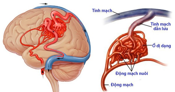
A cavernous hemangioma consists of clusters of small, blood-filled vascular cavities. These represent abnormal formations of capillary vessels, in which the blood-filled spaces are separated by thin septa, giving a characteristic appearance resembling a honeycomb or a mulberry-like structure.
Cavernous hemangiomas are most commonly located within the cerebral hemispheres, although they may also occur in the brainstem, deep brain regions, and, less frequently, in the spinal cord. These lesions are considered benign; however, due to their thin vascular walls, they are prone to recurrent hemorrhage. Although bleeding episodes may be self-limited, the risk of repeated hemorrhage necessitates careful monitoring and should not be underestimated.
Causes of cavernous hemangioma
The exact etiology of cavernous hemangioma remains unclear, and most cases are considered to occur sporadically.
However, accumulating evidence suggests a genetic predisposition. In familial cases, mutations associated with chromosome 7 abnormalities have been identified.
Additionally, from a histopathological perspective, cavernous hemangiomas consist of dilated capillary channels with thin walls lacking normal elastic fibers and smooth muscle components. This structural fragility makes them highly susceptible to hemorrhage, particularly as vascular integrity declines with age.
Clinical manifestations of cavernous hemangioma
The clinical presentation of cavernous hemangioma varies depending on the location and size of the lesion. However, most patients may exhibit the following typical symptoms:
- Seizures and neurological disturbances
- Recurrent intracavernous hemorrhage, with an increasing frequency over time, estimated at approximately 0.5–1% per year
- Headache, nausea, visual disturbances, and drowsiness associated with increased intracranial pressure
Approximately 50% of cases present with symptoms such as headache, seizures, nausea, and limb weakness when hemorrhage occurs within the cavernous hemangioma. The remaining 50% are typically asymptomatic and are incidentally detected during brain imaging studies, such as MRI or CT scans.

Diagnostic approaches for cavernous hemangioma
Cavernous hemangiomas may be symptomatic or asymptomatic; therefore, clinical examination alone is often insufficient for definitive diagnosis.
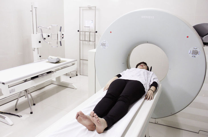
Accurate diagnosis is primarily established through neuroimaging modalities such as brain CT or magnetic resonance imaging (MRI), which allow precise evaluation of the lesion’s size, location, and characteristics.
During the diagnostic process, careful differential diagnosis is essential to distinguish cavernous hemangiomas from other conditions, such as hemorrhagic metastases or low-grade gliomas, in order to avoid misdiagnosis.
Treatment of cavernous hemangioma
Although cavernous hemangioma is a benign condition, it should not be underestimated due to the risk of recurrent hemorrhage. Once diagnosed, patients are advised to seek medical consultation for timely and appropriate management.
Treatment strategies depend on the lesion’s location, size, and severity, and are individualized for each patient.
- Asymptomatic cases: Conservative management is typically recommended, including regular monitoring and annual MRI to assess lesion progression
- Symptomatic cases: Treatment may involve surgical resection or stereotactic radiosurgery (e.g., Gamma Knife), depending on clinical indications and lesion characteristics
- Surgical management is indicated in cases of solitary lesions, hemorrhage located in non-eloquent brain regions that do not compromise neurological function, or cavernous hemangiomas associated with seizures or epilepsy.
- Stereotactic radiosurgery (Gamma Knife) is considered for lesions that have bled and are located in deep or critical areas such as the brainstem, cerebral peduncle, thalamus, or eloquent cortex (motor and sensory regions). This approach is also suitable for patients who are not candidates for surgery, including those with significant comorbidities, advanced age, or those who decline surgical intervention.
Gamma Knife radiosurgery is a modern, minimally invasive technique that utilizes precisely targeted radiation at therapeutic doses to eliminate vascular lesions. This approach is associated with a low risk of complications during and after treatment.
Cavernous hemangioma can be effectively managed when detected at an early stage. Therefore, regular health check-ups and prompt medical evaluation when abnormal symptoms arise are strongly recommended. Early diagnosis and timely treatment not only improve clinical outcomes but also help reduce overall healthcare costs.
Register for consultation and evaluation with specialists at Hong Ngoc General Hospital here:
Note: The information provided in this article by Hong Ngoc General Hospital is for reference purposes only and does not replace professional medical diagnosis or treatment. Patients should not self-medicate. For an accurate assessment of their condition, individuals are advised to visit a medical facility for direct examination, diagnosis, and appropriate treatment planning by qualified physicians.
Follow the official fanpage of Hong Ngoc General Hospital for more useful health information and updates.