Among the four main types of thyroid cancer, papillary thyroid carcinoma is the most common and also the one with the highest treatment success rate. However, if not detected and treated in a timely manner, papillary thyroid carcinoma may progress to severe complications and pose serious risks to health.
What is papillary thyroid carcinoma?
Papillary thyroid carcinoma (PTC) is a type of malignant tumor that develops within the thyroid gland, arising from abnormal cancerous cells in thyroid tissue.
This is the most common form of thyroid cancer, accounting for up to 90% of all cases. The condition most frequently affects middle-aged individuals, with the average age of diagnosis around 50. Women are three times more likely to develop papillary thyroid carcinoma than men.
Symptoms of papillary thyroid carcinoma

Although papillary thyroid carcinoma has a high treatment success rate, it is often difficult to detect at an early stage because it usually causes no symptoms initially. As the disease progresses, patients may experience the following manifestations:
- Enlargement of the thyroid gland: One of the main signs of papillary thyroid carcinoma. The thyroid may become noticeably larger and feel firmer on palpation.
- Difficulty swallowing or breathing, often accompanied by pain: An enlarged thyroid can compress the esophagus and trachea, leading to dysphagia, shortness of breath, and discomfort.
- Hoarseness or voice changes: Tumor growth within the thyroid may place pressure on the vocal cords, resulting in hoarseness or alteration of the voice.
- Cervical lymph node metastasis: Commonly involving level VI lymph nodes, which are typically firm, painless, and mobile when not yet invasive. Notably, about one-third of patients experience lymphatic spread beyond the neck, with metastasis to the lungs.
Causes of papillary thyroid carcinoma
The exact cause of papillary thyroid carcinoma (PTC) remains unclear. However, according to reputable medical literature, several factors are known to increase the risk of developing this disease:
- Genetic predisposition: Certain inherited genetic alterations can contribute to the development of PTC, such as RET-related syndromes (multiple endocrine neoplasia – MEN), thyroglobulin gene mutations, SDHx mutations, PTEN mutations, and other variants. In fact, approximately 5% of papillary thyroid carcinoma cases are familial.
- Environmental factors: Exposure to environmental triggers such as radiation, air pollution, iodine-containing drinking water, or assisted reproductive technologies may increase the risk of PTC.
- Thyroid lesions and tumors: Structural abnormalities or tumors of the thyroid may raise the likelihood of malignancy. Additionally, other thyroid conditions such as neuroendocrine thyroid tumors and Hürthle cell thyroid disease are associated with an elevated risk of papillary thyroid carcinoma.
Is papillary thyroid carcinoma dangerous?
Patients under 55 years of age usually have a good prognosis if the disease is diagnosed at an early stage. However, about 10% of patients with papillary thyroid carcinoma present with cervical lymph node metastasis on the same side as the tumor.
In recent decades, the incidence of papillary thyroid carcinoma has increased significantly, as many patients incidentally discover small tumors during examinations such as neck ultrasound, MRI, CT, or PET scans.

Therefore, regular thyroid examinations play a crucial role in detecting papillary thyroid carcinoma, as the disease often shows no specific symptoms in its early stages. Once detected, delaying treatment may result in missing the optimal window for effective therapy.
To understand your condition clearly, you should seek professional advice from endocrinologists. Schedule an appointment with our Thyroid–Endocrinology specialists via hotline 0911.858.626 or register [HERE].
Diagnostic methods for papillary thyroid carcinoma
Cytological diagnosis (Fine-needle aspiration – FNA)
This is the primary diagnostic method used to confirm papillary thyroid carcinoma, with an accuracy rate of up to 95%.
The procedure is performed by inserting a fine needle into the thyroid nodule to collect cell samples. These samples are then analyzed in the laboratory to detect any abnormal thyroid cells suggestive of malignancy.

Radioiodine thyroid scintigraphy (I-131 scan) Thyroid scintigraphy assists in detecting papillary thyroid carcinoma, as malignant cells usually have low iodine uptake and often appear as hypofunctioning (“cold”) areas on the scan. In addition, whole-body scintigraphy with I-131 can help identify distant metastases to other organs.
Imaging studies These include ultrasound, CT, and MRI. Ultrasound is the most commonly used imaging modality to evaluate thyroid nodules. CT and MRI may be employed to determine the exact size and location of the tumor and to detect other structural abnormalities or evidence of disease spread.
Treatment of papillary thyroid carcinoma
Case: Tumors >4 cm or with metastasis
In these cases, patients are typically indicated for surgery to remove the thyroid gland, either partially or completely. Following surgery, adjuvant radioiodine therapy (I-131) is administered at appropriate doses to eradicate any residual thyroid tissue. This therapy may be repeated every 6 to 12 months to ensure complete ablation of remaining thyroid tissue. After surgery and radioiodine therapy, patients are required to take thyroid-stimulating hormone (TSH) suppressive therapy with levothyroxine (T4), since TSH can stimulate the growth of any residual papillary thyroid carcinoma cells.
Case: Tumors <4 cm confined to one thyroid lobe
For tumors of this size and location, experts often recommend lobectomy (removal of one thyroid lobe) or isthmusectomy (removal of the thyroid isthmus). Surgery remains the most effective treatment option. Postoperatively, patients are usually prescribed low-dose TSH suppression therapy to reduce the risk of recurrence and to suppress any microscopic residual papillary thyroid carcinoma.
Papillary thyroid carcinoma <1.5 cm without evidence of metastasis
In patients with papillary thyroid carcinoma smaller than 1.5 cm, with no abnormal lymph nodes or signs of metastasis on diagnostic evaluation, active surveillance combined with medical therapy may be an appropriate management approach.
Patients must attend regular follow-up visits as scheduled and strictly adhere to their physician’s recommendations to prevent cancer cell progression, which could negatively affect health outcomes and treatment effectiveness.
Reader may also be interested in:
- Thyroid cancer – What causes it? Endocrinology experts warn of five potential complications of thyroid cancer
- Thyroid nodules: Causes, symptoms, and treatment options
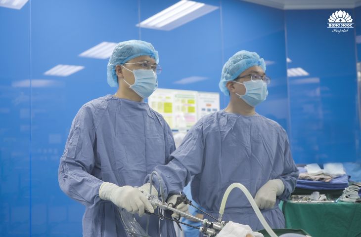
Trusted center for thyroid cancer diagnosis and treatment
At Hong Ngoc General Hospital, patients are offered advanced diagnostic tests and individualized treatment plans tailored to their specific condition.
With a team of highly experienced physicians and state-of-the-art medical equipment, the Thyroid Center at Hong Ngoc General Hospital performs more than 1,000 thyroid surgeries each year, meeting the full spectrum of patient care needs.
The center is capable of carrying out highly complex surgical procedures, including transoral endoscopic thyroidectomy for thyroid cancer. In addition to conventional open and laparoscopic surgery, Hong Ngoc is among the few hospitals in Vietnam able to apply this advanced scar-free endoscopic technique and has successfully treated hundreds of thyroid cancer patients.
Contact Information Department of Endocrinology – Hong Ngoc General Hospital – Address: No. 8 Chau Van Liem, Nam Tu Liem, Hanoi – Hotline: 0911 858 626
Note: The information provided in this article by Hong Ngoc General Hospital is for reference only and is not a substitute for medical diagnosis or treatment. For an accurate diagnosis and effective treatment plan, patients should visit reputable medical facilities for direct consultation and examination by physicians.
Follow the official Hong Ngoc General Hospital fanpage to receive more useful health information and updates.