Urolithiasis is a common condition, currently affecting an estimated 2–12% of the Vietnamese population. But can urinary stones lead to dangerous complications?
Urolithiasis includes kidney stones, bladder stones, ureteral stones, and urethral stones. Below is essential information on urolithiasis and its treatment.
The urinary system and causes of urolithiasis
The urinary system is located within the abdominal cavity and serves as the body’s natural filtration mechanism. Its main function is to filter the blood and excrete waste products such as urea, uric acid, and ammonia through urine.
The urinary system consists of:
Two kidneys, responsible for blood filtration and urine production
Two ureters, transporting urine from the kidneys to the bladder
One bladder, serving as a reservoir for urine before excretion
One urethra, carrying urine from the bladder to the outside of the body
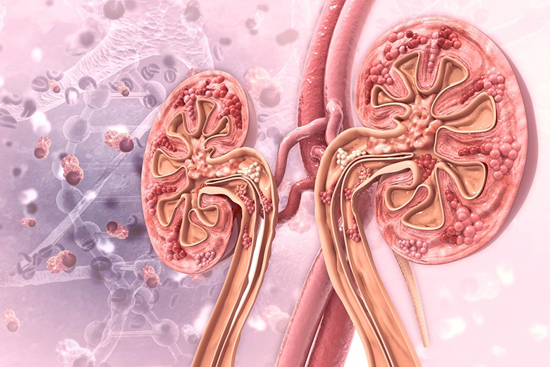
Illustration of the urinary system
Urolithiasis develops when crystals in the urine aggregate and deposit under favorable conditions such as:
Reduced urinary flow or altered urinary pH
Urinary tract infections
Congenital or acquired abnormalities of the urinary tract
Other factors that increase the risk of urinary stone formation include:
Dehydration due to insufficient fluid intake
Diets high in protein, sodium (salt), and sugar
Gastrointestinal diseases or previous surgeries that alter digestion and absorption, leading to changes in calcium and fluid balance and increasing stone-forming substances in the urine
Classification of urolithiasis
Urolithiasis can be classified in different ways, most commonly by their chemical composition or by their anatomical location.
Classification by chemical composition
Inorganic stones
Calcium oxalate stones: Typically black, spiculated, with high radiopacity.
Calcium phosphate stones: Pale yellow or opaque white, brittle.
Calcium carbonate stones: Chalky white, soft, and fragile.
Organic stones
Urate stones: Milky white, opaque, may be radiolucent, soft, and prone to recurrence.
Cystine stones: Pale yellow, soft, often recurrent.
Struvite stones: Yellowish-white, usually associated with urinary tract infections caused by Proteus species.
In Vietnam, the majority of urinary stone cases are inorganic, with calcium oxalate stones accounting for over 80%. These stones often exist as mixed forms containing multiple chemical components.
Classification by anatomical location
Kidney stones (40%): Including renal pelvis stones, calyceal stones, pelvicalyceal stones, staghorn stones, and partial staghorn stones.
Ureteral stones (28%)
Bladder stones (26%)
Urethral stones (4%)
Characteristics of urolithiasis
Number of stones: The number of urolithiasis can vary, ranging from a single stone to hundreds. Urolithiasis are most often solitary before treatment (lithotripsy).
Size of stones: Urinary stones can measure from a few millimeters to several centimeters. Stones located in the renal pelvis are usually larger than those found elsewhere in the urinary tract.
Shape of stones: The morphology of urolithiasis varies depending on their type. Kidney stones are often staghorn-shaped or resemble parrot beaks, while ureteral stones are usually elongated, following the course of the ureter.
If you suspect urolithiasis and wish to receive medical advice on the most suitable treatment options, you may schedule a consultation with our specialists via hotline 0912.002.131 or leave your information [HERE].
How urolithiasis damage the urinary system?
Obstruction caused by stones:
Stones located in the renal pelvis may cause partial or complete obstruction depending on their size. This leads to progressive dilatation of the renal parenchyma, thinning of renal tissue, enlargement of the renal pelvis and calyces, and eventually renal atrophy and loss of kidney function.
Stones in the ureter may obstruct urinary flow, gradually leading to ureteral dilatation, impaired peristalsis, and fibrosis.
Large bladder stones that cannot pass through the urethra may cause urinary retention, ascending infections, and severe complications such as vesicovaginal fistula or bladder shrinkage. In cases where stones obstruct both kidneys and ureters simultaneously, acute renal failure may occur, which is a medical emergency.
Injury caused by friction Hard and spiculated stones, such as calcium oxalate stones, can tear or scratch the lining of the renal pelvis and ureter as they move, creating favorable conditions for urinary tract infection. Friction may also cause bleeding within the urinary tract, accelerate fibrosis of the renal parenchyma and ureter, and lead to ureteral stricture, thereby worsening obstruction. The eventual outcome is progressive renal failure.
Urinary tract infection (UTI) Infections may develop as a result of obstruction or friction between the stone and the lining of the renal pelvis or ureter. This can cause edema, ulceration, and gradual fibrosis of the mucosa, ultimately leading to renal failure. In addition, urinary tract infections produce bacterial debris, white blood cells, and epithelial cells, which can precipitate and crystallize within the urinary tract. In the presence of stones as foreign bodies, this process is further facilitated, promoting the formation of new stones.
Kidney failure caused by urinary stones
Symptoms of urolithiasis to be aware of
The clinical presentation of urolithiasis varies depending on the location of the stone. Some patients may remain completely asymptomatic, with stones detected incidentally during routine examinations, while others experience severe and noticeable symptoms.
The most common symptom is flank or lower back pain, considered the hallmark of urolithiasis. This may present as:
A dull, persistent ache, or sudden, severe colicky pain radiating to the front of the abdomen or the groin and genital region.
Renal colic caused by urolithiasis complications
Abnormal urination: Patients with urolithiasis may experience painful urination (dysuria), interrupted urination (stopping and starting midstream, then resuming when changing position), difficulty urinating, complete urinary retention, or cloudy urine.
Fever: A common sign in patients who have developed a urinary tract infection as a complication of urinary stones.
Diagnostic methods for urolithiasis
Ultrasound
Ultrasound is one of the most commonly used diagnostic methods, allowing evaluation of the location, size, and number of stones as well as the degree of damage to the kidneys and bladder by assessing renal parenchymal thickness or detecting hematoma. Because the procedure is simple and quick, ultrasound is suitable for children and patients who may have difficulty cooperating with other diagnostic techniques. However, to determine the type of stone, additional diagnostic methods may be required.
X-ray
X-ray is a diagnostic technique that uses penetrating X-rays with high sensitivity, providing valuable information to guide effective treatment planning.
Plain abdominal X-ray (KUB): A basic test capable of detecting approximately 90% of stones, particularly radiopaque types such as struvite, calcium phosphate, and calcium oxalate. It provides useful information about the shape, size, number, and location of stones.
Intravenous urography (IVU/IVP): Involves injection of contrast medium to visualize the urinary tract. This method helps detect both radiolucent and radiopaque stones, assess the degree of hydronephrosis, ureteral dilation, and evaluate bladder mucosal damage.
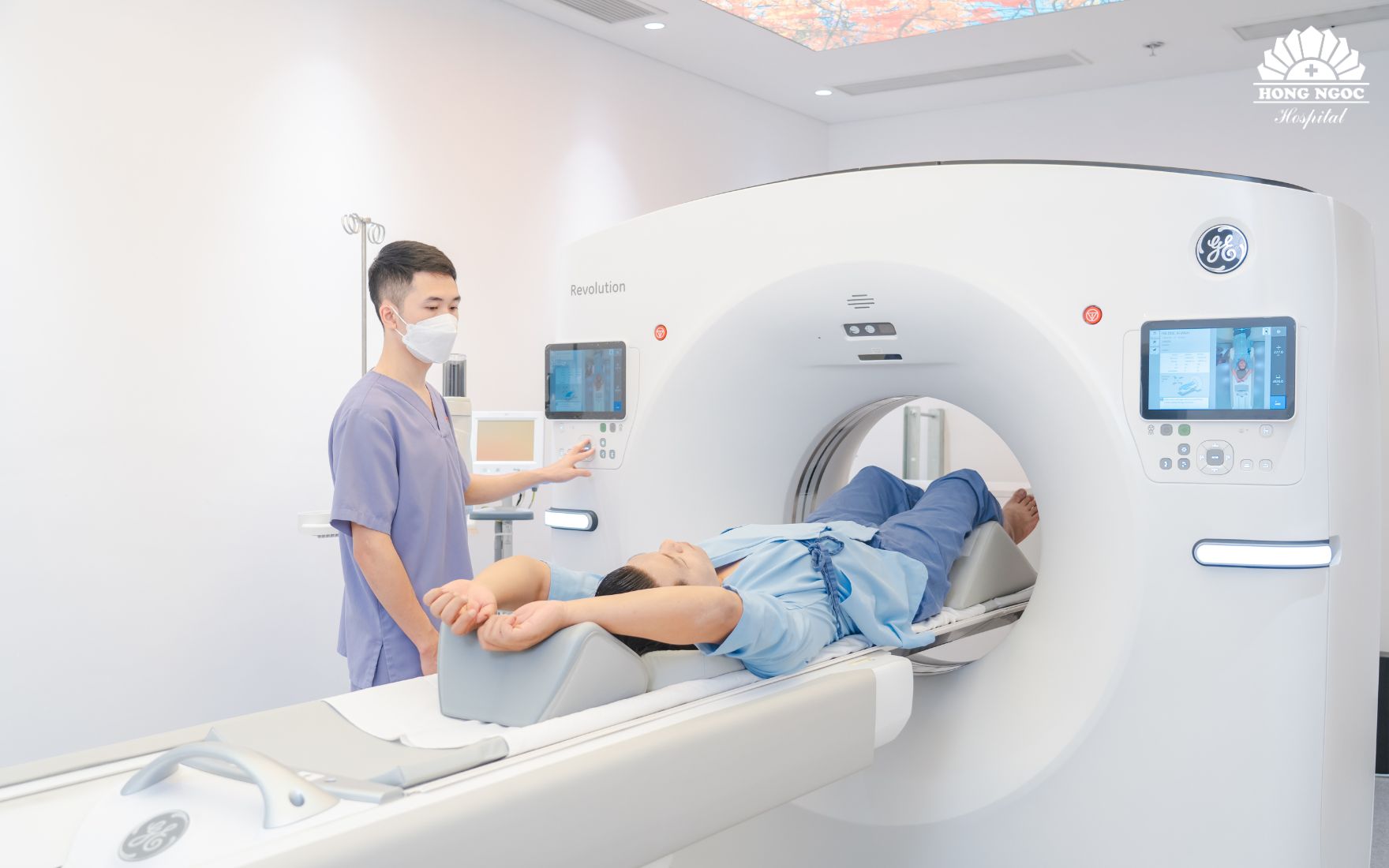
Computed tomography (CT scan)
Multislice CT imaging not only facilitates accurate diagnosis of kidney stone–related conditions but also allows physicians to evaluate renal pathology, renal function, and surrounding tissues and organs with nearly 100% precision.
Importantly, this advanced imaging technique can also differentiate kidney stones from other abnormalities such as urinary tract tumors or congenital malformations, ensuring accurate diagnosis and appropriate treatment planning.
CT scan at Hong Ngoc General Hospital
Blood and urine tests
A complete blood count (CBC) showing decreased red blood cells and elevated white blood cells can help diagnose urinary tract infection and assess damage to the urinary system.
Urinalysis to detect red blood cells, white blood cells, or oxalate and phosphate crystals (components commonly found in stones) helps evaluate their presence.
In addition, biochemical tests measuring urea and creatinine levels help assess kidney failure, while radioisotope studies or urine culture are used to evaluate urinary tract infection.
Urolithiasis can be completely treated with a variety of methods, ranging from medical management to advanced surgical interventions.
Medical treatment
This approach is effective in cases where stones are smaller than 5 mm and have not caused complications. The treatment plan is tailored by the physician based on the stone’s location, size, composition, and the extent of urinary tract damage.
Advanced stone fragmentation techniques
Modern minimally invasive techniques have significantly improved treatment outcomes, helping patients avoid open surgery, which carries higher risks of complications and longer recovery times.
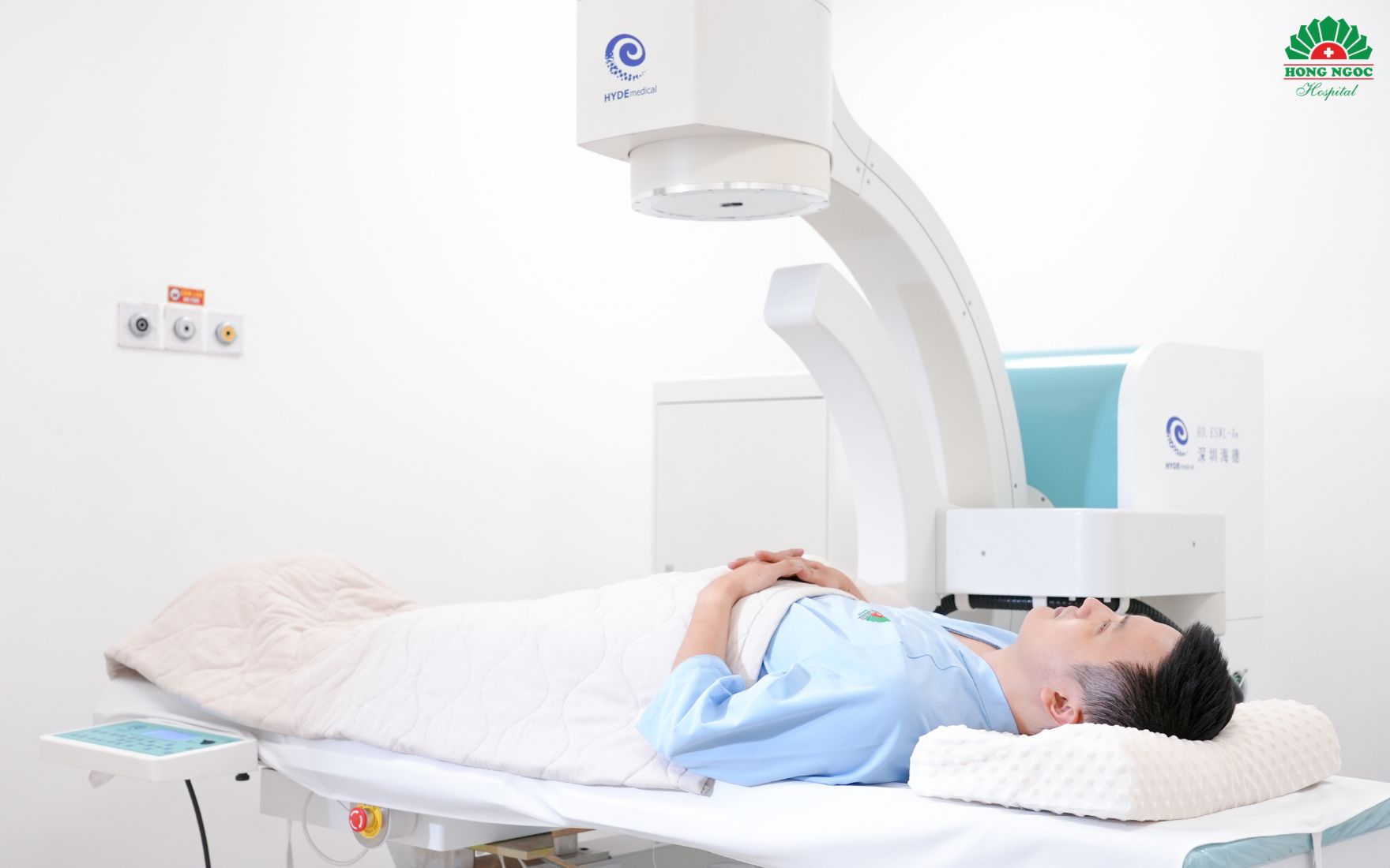
Extracorporeal shock wave lithotripsy (ESWL)
This method uses shock waves directed from outside the body to break kidney stones or upper ureteral stones into tiny fragments. These fragments are then naturally expelled through the urinary tract without the need for surgical intervention.
With extracorporeal shock wave lithotripsy (ESWL), hospitalization is not required.
Percutaneous laser lithotripsy Instead of open surgery to remove large kidney stones or stones located deep within the renal calyces, percutaneous laser lithotripsy provides a breakthrough solution. The entire procedure is performed through a very small tract, only about 5 mm in size. A single, shortest-access tract is created to reach the stone. Through this tract, a nephroscope is introduced, and a high-energy laser is used to fragment the stones—including large or very hard ones—into tiny pieces. This method minimizes residual fragments while preserving renal parenchyma to the greatest extent possible.
Percutaneous laser lithotripsy successfully removed hundreds of stones in a 67-year-old patient.
Retrograde ureteroscopic laser lithotripsy This technique also employs high-power laser energy to break stones into small fragments for removal. However, instead of entering through the kidney, the laser source is delivered via a ureteroscope introduced naturally in a retrograde fashion through the urethra, bladder, and up into the ureter or renal pelvis. High-definition endoscopic systems provide clear, detailed visualization, helping physicians identify stones even in complex locations and minimize the risk of residual fragments.
Depending on the stone’s location, physicians will select the appropriate approach:
Rigid ureteroscope: Best suited for stones in the distal ureter or bladder.
Flexible ureteroscope: Indicated for kidney stones or stones in the upper ureter.
Where to seek care for urolithiasis?
In Vietnam, urolithiasis are often detected incidentally or only after significant symptoms have developed. To prevent stones from enlarging and causing serious complications, individuals are advised to undergo regular health check-ups and seek medical evaluation at the first signs of urinary stone disease.
Hong Ngoc General Hospital is recognized as a trusted center for the diagnosis and treatment of urinary tract disorders, particularly urolithiasis. With modern facilities, international-standard operating theaters, and a team of highly skilled, experienced physicians, the hospital has successfully mastered advanced urological surgical techniques that are both safe and effective.
Patients receive detailed counseling on their condition at the Urology Clinic – Hong Ngoc General Hospital.
Patients are examined and treated directly by a team of highly qualified, experienced urologists who provide thorough consultation to help each individual choose the most effective and suitable stone treatment method. Leading the urology expert council is Dr. Trinh Minh Thanh, MSc, Specialist Level II, with outstanding credentials:
Over 30 years of clinical experience at Bach Mai Hospital and Viet Duc Hospital
Certified in Urological Surgery – Kobe University, Japan; certified in Laparoscopic Surgery – United States
Performed thousands of complex urological surgeries, including cases of urinary stones, cancers, urinary tract malformations, and prostate tumors
**Note: The information provided in this article by Hong Ngoc General Hospital is for reference only and is not a substitute for professional medical diagnosis or treatment. Patients should not self-medicate. To obtain an accurate diagnosis and appropriate treatment plan, please visit a hospital for direct examination and consultation with a physician.
Follow the official fanpage of Hong Ngoc General Hospital [HERE] to receive more useful health information and updates on the hospital’s special promotions.
Urolithiasis is a common condition, currently affecting an estimated 2–12% of the Vietnamese population. But can urinary stones lead to dangerous complications?
Urolithiasis includes kidney stones, bladder stones, ureteral stones, and urethral stones. Below is essential information on urolithiasis and its treatment.
The urinary system and causes of urolithiasis
The urinary system is located within the abdominal cavity and serves as the body’s natural filtration mechanism. Its main function is to filter the blood and excrete waste products such as urea, uric acid, and ammonia through urine.
The urinary system consists of:
Two kidneys, responsible for blood filtration and urine production
Two ureters, transporting urine from the kidneys to the bladder
One bladder, serving as a reservoir for urine before excretion
One urethra, carrying urine from the bladder to the outside of the body
Illustration of the urinary system
Urolithiasis develops when crystals in the urine aggregate and deposit under favorable conditions such as:
Reduced urinary flow or altered urinary pH
Urinary tract infections
Congenital or acquired abnormalities of the urinary tract
Other factors that increase the risk of urinary stone formation include:
Dehydration due to insufficient fluid intake
Diets high in protein, sodium (salt), and sugar
Gastrointestinal diseases or previous surgeries that alter digestion and absorption, leading to changes in calcium and fluid balance and increasing stone-forming substances in the urine
Classification of urolithiasis
Urolithiasis can be classified in different ways, most commonly by their chemical composition or by their anatomical location.
Classification by chemical composition
Inorganic stones
Calcium oxalate stones: Typically black, spiculated, with high radiopacity.
Calcium phosphate stones: Pale yellow or opaque white, brittle.
Calcium carbonate stones: Chalky white, soft, and fragile.
Organic stones
Urate stones: Milky white, opaque, may be radiolucent, soft, and prone to recurrence.
Cystine stones: Pale yellow, soft, often recurrent.
Struvite stones: Yellowish-white, usually associated with urinary tract infections caused by Proteus species.
In Vietnam, the majority of urinary stone cases are inorganic, with calcium oxalate stones accounting for over 80%. These stones often exist as mixed forms containing multiple chemical components.
Classification by anatomical location
Kidney stones (40%): Including renal pelvis stones, calyceal stones, pelvicalyceal stones, staghorn stones, and partial staghorn stones.
Ureteral stones (28%)
Bladder stones (26%)
Urethral stones (4%)
Characteristics of urolithiasis
Number of stones: The number of urolithiasis can vary, ranging from a single stone to hundreds. Urolithiasis are most often solitary before treatment (lithotripsy).
Size of stones: Urinary stones can measure from a few millimeters to several centimeters. Stones located in the renal pelvis are usually larger than those found elsewhere in the urinary tract.
Shape of stones: The morphology of urolithiasis varies depending on their type. Kidney stones are often staghorn-shaped or resemble parrot beaks, while ureteral stones are usually elongated, following the course of the ureter.
If you suspect urolithiasis and wish to receive medical advice on the most suitable treatment options, you may schedule a consultation with our specialists via hotline 0912.002.131 or leave your information [HERE].
How urolithiasis damage the urinary system?
Obstruction caused by stones:
Stones located in the renal pelvis may cause partial or complete obstruction depending on their size. This leads to progressive dilatation of the renal parenchyma, thinning of renal tissue, enlargement of the renal pelvis and calyces, and eventually renal atrophy and loss of kidney function.
Stones in the ureter may obstruct urinary flow, gradually leading to ureteral dilatation, impaired peristalsis, and fibrosis.
Large bladder stones that cannot pass through the urethra may cause urinary retention, ascending infections, and severe complications such as vesicovaginal fistula or bladder shrinkage. In cases where stones obstruct both kidneys and ureters simultaneously, acute renal failure may occur, which is a medical emergency.
Injury caused by friction Hard and spiculated stones, such as calcium oxalate stones, can tear or scratch the lining of the renal pelvis and ureter as they move, creating favorable conditions for urinary tract infection. Friction may also cause bleeding within the urinary tract, accelerate fibrosis of the renal parenchyma and ureter, and lead to ureteral stricture, thereby worsening obstruction. The eventual outcome is progressive renal failure.
Urinary tract infection (UTI) Infections may develop as a result of obstruction or friction between the stone and the lining of the renal pelvis or ureter. This can cause edema, ulceration, and gradual fibrosis of the mucosa, ultimately leading to renal failure. In addition, urinary tract infections produce bacterial debris, white blood cells, and epithelial cells, which can precipitate and crystallize within the urinary tract. In the presence of stones as foreign bodies, this process is further facilitated, promoting the formation of new stones.
Kidney failure caused by urinary stones
Symptoms of urolithiasis to be aware of
The clinical presentation of urolithiasis varies depending on the location of the stone. Some patients may remain completely asymptomatic, with stones detected incidentally during routine examinations, while others experience severe and noticeable symptoms.
The most common symptom is flank or lower back pain, considered the hallmark of urolithiasis. This may present as:
A dull, persistent ache, or sudden, severe colicky pain radiating to the front of the abdomen or the groin and genital region.
Renal colic caused by urolithiasis complications
Abnormal urination: Patients with urolithiasis may experience painful urination (dysuria), interrupted urination (stopping and starting midstream, then resuming when changing position), difficulty urinating, complete urinary retention, or cloudy urine.
Fever: A common sign in patients who have developed a urinary tract infection as a complication of urinary stones.
Diagnostic methods for urolithiasis
Ultrasound
Ultrasound is one of the most commonly used diagnostic methods, allowing evaluation of the location, size, and number of stones as well as the degree of damage to the kidneys and bladder by assessing renal parenchymal thickness or detecting hematoma. Because the procedure is simple and quick, ultrasound is suitable for children and patients who may have difficulty cooperating with other diagnostic techniques. However, to determine the type of stone, additional diagnostic methods may be required.
X-ray
X-ray is a diagnostic technique that uses penetrating X-rays with high sensitivity, providing valuable information to guide effective treatment planning.
Plain abdominal X-ray (KUB): A basic test capable of detecting approximately 90% of stones, particularly radiopaque types such as struvite, calcium phosphate, and calcium oxalate. It provides useful information about the shape, size, number, and location of stones.
Intravenous urography (IVU/IVP): Involves injection of contrast medium to visualize the urinary tract. This method helps detect both radiolucent and radiopaque stones, assess the degree of hydronephrosis, ureteral dilation, and evaluate bladder mucosal damage.
Computed tomography (CT scan)
Multislice CT imaging not only facilitates accurate diagnosis of kidney stone–related conditions but also allows physicians to evaluate renal pathology, renal function, and surrounding tissues and organs with nearly 100% precision.
Importantly, this advanced imaging technique can also differentiate kidney stones from other abnormalities such as urinary tract tumors or congenital malformations, ensuring accurate diagnosis and appropriate treatment planning.
CT scan at Hong Ngoc General Hospital
Blood and urine tests
A complete blood count (CBC) showing decreased red blood cells and elevated white blood cells can help diagnose urinary tract infection and assess damage to the urinary system.
Urinalysis to detect red blood cells, white blood cells, or oxalate and phosphate crystals (components commonly found in stones) helps evaluate their presence.
In addition, biochemical tests measuring urea and creatinine levels help assess kidney failure, while radioisotope studies or urine culture are used to evaluate urinary tract infection.
Urolithiasis can be completely treated with a variety of methods, ranging from medical management to advanced surgical interventions.
Medical treatment
This approach is effective in cases where stones are smaller than 5 mm and have not caused complications. The treatment plan is tailored by the physician based on the stone’s location, size, composition, and the extent of urinary tract damage.
Advanced stone fragmentation techniques
Modern minimally invasive techniques have significantly improved treatment outcomes, helping patients avoid open surgery, which carries higher risks of complications and longer recovery times.
Extracorporeal shock wave lithotripsy (ESWL)
This method uses shock waves directed from outside the body to break kidney stones or upper ureteral stones into tiny fragments. These fragments are then naturally expelled through the urinary tract without the need for surgical intervention.
With extracorporeal shock wave lithotripsy (ESWL), hospitalization is not required.
Percutaneous laser lithotripsy Instead of open surgery to remove large kidney stones or stones located deep within the renal calyces, percutaneous laser lithotripsy provides a breakthrough solution. The entire procedure is performed through a very small tract, only about 5 mm in size. A single, shortest-access tract is created to reach the stone. Through this tract, a nephroscope is introduced, and a high-energy laser is used to fragment the stones—including large or very hard ones—into tiny pieces. This method minimizes residual fragments while preserving renal parenchyma to the greatest extent possible.
Percutaneous laser lithotripsy successfully removed hundreds of stones in a 67-year-old patient.
Retrograde ureteroscopic laser lithotripsy This technique also employs high-power laser energy to break stones into small fragments for removal. However, instead of entering through the kidney, the laser source is delivered via a ureteroscope introduced naturally in a retrograde fashion through the urethra, bladder, and up into the ureter or renal pelvis. High-definition endoscopic systems provide clear, detailed visualization, helping physicians identify stones even in complex locations and minimize the risk of residual fragments.
Depending on the stone’s location, physicians will select the appropriate approach:
Rigid ureteroscope: Best suited for stones in the distal ureter or bladder.
Flexible ureteroscope: Indicated for kidney stones or stones in the upper ureter.
Where to seek care for urolithiasis?
In Vietnam, urolithiasis are often detected incidentally or only after significant symptoms have developed. To prevent stones from enlarging and causing serious complications, individuals are advised to undergo regular health check-ups and seek medical evaluation at the first signs of urinary stone disease.
Hong Ngoc General Hospital is recognized as a trusted center for the diagnosis and treatment of urinary tract disorders, particularly urolithiasis. With modern facilities, international-standard operating theaters, and a team of highly skilled, experienced physicians, the hospital has successfully mastered advanced urological surgical techniques that are both safe and effective.
Patients receive detailed counseling on their condition at the Urology Clinic – Hong Ngoc General Hospital.
Patients are examined and treated directly by a team of highly qualified, experienced urologists who provide thorough consultation to help each individual choose the most effective and suitable stone treatment method. Leading the urology expert council is Dr. Trinh Minh Thanh, MSc, Specialist Level II, with outstanding credentials:
Over 30 years of clinical experience at Bach Mai Hospital and Viet Duc Hospital
Certified in Urological Surgery – Kobe University, Japan; certified in Laparoscopic Surgery – United States
Performed thousands of complex urological surgeries, including cases of urinary stones, cancers, urinary tract malformations, and prostate tumors
**Note: The information provided in this article by Hong Ngoc General Hospital is for reference only and is not a substitute for professional medical diagnosis or treatment. Patients should not self-medicate. To obtain an accurate diagnosis and appropriate treatment plan, please visit a hospital for direct examination and consultation with a physician.
Follow the official fanpage of Hong Ngoc General Hospital [HERE] to receive more useful health information and updates on the hospital’s special promotions.
Để lại câu hỏi của bạn để nhận được giải đáp từ các bác sĩ của Hồng Ngọc
Lorem ipsum dolor
Lorem ipsum dolor sit amet, consectetur adipiscing elit. Mauris odio lectus, pretium faucibus nisi eu, accumsan consectetur orci. In blandit vehicula nisl, vel lacinia ligula finibus a. Donec fermentum rhoncus
Lorem ipsum dolor sit amet, consectetur adipiscing elit. Mauris odio lectus, pretium faucibus nisi eu, accumsan consectetur orci. In blandit vehicula nisl, vel lacinia ligula finibus a. Donec fermentum rhoncus
Lorem ipsum dolor sit amet, consectetur adipiscing elit. Mauris odio lectus, pretium faucibus nisi eu, accumsan consectetur orci. In blandit vehicula nisl, vel lacinia ligula finibus a. Donec fermentum rhoncus
Lorem ipsum dolor sit amet, consectetur adipiscing elit. Mauris odio lectus, pretium faucibus nisi eu, accumsan consectetur orci. In blandit vehicula nisl, vel lacinia ligula finibus a. Donec fermentum rhoncus
Lorem ipsum dolor sit amet, consectetur adipiscing elit. Mauris odio lectus, pretium faucibus nisi eu, accumsan consectetur orci. In blandit vehicula nisl, vel lacinia ligula finibus a. Donec fermentum rhoncus